|
The author's abstract of Derkach A.A. masters works
Plan:
Introduction
- Urgency of a theme.
- The description of a subject domain.
- Statement of a problem.
- Similar domestic and foreign development.
- Problems, the purposes and expected results of masters works.
The conclusion.
The list of sources.
Introduction
Researches of preparations under a microscope it is widely applied in modern medicine. Especially it touches the researches connected with cells of fabrics of the person (blood, a lymph, mucous membranes, etc.) Development of modern means of input of the image from a microscope (inexpensive photo-and videocameras) in a computer has allowed to process images with use of various methods of morphological processing. As well as any optical device, a microscope demands some exact tincture of focus. In most cases this adjustment is carried out by the operator of a research complex. It is necessary it manually, rotating adjusting screws to establish focus so that it was possible to receive as much as possible precise image. This process have a number of lacks:
· Expenses of time (frequently more time it is spent for adjustment of a microscope, than for the analysis of the received image)
· High probability of inexact adjustment (especially at operators with the lowered sight or which use glasses)
· Application of manual skills there, where probably application of automatic system
· the Raised loading on the visual device of the operator
Thus the operator makes focusing for the image. In practice it is necessary to be focused or on any object on the image (for example, a cellular membrane) or on a part of this object more often. At manual fine tuning a microscope it is very difficult to achieve sufficient image sharpness in area necessary for research.
[Top]
Urgency of a theme.
In connection with presence of lacks of manual focusing of the image designing and creation of the automated subsystem of focusing of a microscope is an actual problem{task}. Practical importance of the given subsystem will be defined{determined} by an opportunity of acceleration of process of the analysis and processing of the medical images received from a microscope. In turn it will allow to accelerate process of treatment or definition of the diagnosis of the patient.
[Top]
The description of a subject domain.
Object of research are images of blood cells of the person. Images receive in vitro - in normal conditions. They are applied to the analysis and forecasting of the processes occuring to blood cells with the purpose of increase of efficiency of diagnostics and treatment. In the given work I shall investigate images of such blood cells, as Neutrofils. Neutrofils [3] - the most numerous version white blood cell, they make 50-75 % of all leukocytes. Are named so for appearance cytoplasm granules at painting on Hymza. Depending on a degree of a maturity and the form of a kernel in peripheral blood allocate stickkernel (younger) and segmentkernel (mature) neutrofils. Younger cells Neutrofils of some - young (metamielocits), mielocits, promielocits - appear in peripheral blood in case of a pathology and are the certificate of stimulation of formation of cells of this kind. Their basic function - protection against infections by hemotaxic (the directed movement to stimulating agents) and fagocitose (absorption and digestion) alien microorganisms. [2] in particular us the processes occuring with Neutrofils will interest:
· Process of an attachment
· Fagositose
The example of the image Neutrofils, received from a microscope, is presented in figure 1:
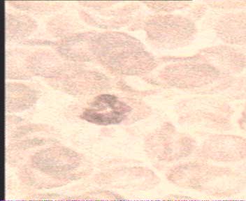
Fig. 1 – Neutrofils image, received from a microscope
[Наверх]
Statement of a problem.
Development of a subsystem of automatic focusing will be spent on the basis of research laboratory of scientific research institute of medical problems of family. In laboratory the usual light microscope is established. The eyepiece of a microscope is interfaced to an analog videocamera, the signal from which acts on an input of a videocard of a computer. Using special program maintenance for capture of images, receive photos of cellular structures in the necessary increase. The general scheme of this complex in its present kind is presented in following figure:
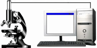
Fig. 2 - the Scheme of a complex for processing the images received from a microscope
(Animation, 5 frames,repeat,0.5 sec for frame)
The images received from a microscope, require additional focusing. The opportunity to execute it in two operating modes is necessary:
1) Automatic in a mode of real time - when the image is grasped directly from the chamber and after processing on inputs of the operating mechanism of adjustment the signal for fine tuning focus moves. After that there is a direct capture and preservation of the image of necessary quality.
2) Processing of images after capture - in the program opens a series of images of which by the analysis the most focused image which then and is used gets out.
Thus for focusing the operator is touched object on the image or a part of this object. It is the important difference of projected system in comparison with earlier created development where focusing was spent under the image as a whole. As a result of performance master works it is necessary to choose on the basis of the analysis of various methods the optimal on speed and accuracy, to design and realize a computer subsystem of automatic focusing of a microscope with an opportunity of work in two above-stated modes.
[Top]
Similar domestic and foreign development
In problem of automatic focusing and definition focusing images is engaged a lot of domestic (I mean scientists it from the CIS countries) and foreign scientists. The set of clauses, scientific works and books is devoted to the given problem, and also a plenty of researches is lead. Also set of the development based on a method of focusing, constructions of three-dimensional model of a surface and other are applied to the analysis of images.. Below I shall result, in my opinion, most significant of them.
1) Morphological methods of interpretation of measurements of a relief of a surface by means of an optical microscope.[2]
2) AutoMontage PRO - the software for work with three-dimensional images in microscopy.[4]
3) analySIS work - the basic package of reception and archiving images from Olympus for a science of materials.[5]
4) Digital microscope KH-3000 firms HIROX (Japan)
[Top]
Problems, the purposes and expected results of master works.
In master work the problem{ of the analysis and a choice optimum both on productivity, and on quality of result, a method of definition and automatic focusing on object on the image is solved.
As a result of performance master works scientific research of methods should be executed. After a choice of an optimum method it is necessary to realize a software package for processing images by this method.
The ready software package should be tested on a set of real data. Results of experiment will allow to judge reliability or unauthenticity results of work of a software package.
[Top]
The conclusion
As a result of performance master works have been investigated various methods of autofocusing of the image received from a microscope. As the basic methods for master works have been chosen following methods:
1.A method of dispersions of gradation grey.[1]
2.A method of size of a gradient. [1]
3.The methods based on the second derivatives.[1]
Have been carried out practical researches of methods, their program realization is created. The software package using these methods, has successfully coped with test sample of images.
[Top]
The list of sources:
1. Diatom autofocusing in brighteld microscopy: a comparative study
Link:http://www.iv.optica.csic.es/papers/icpr2k.pdf
2. "Morphological methods of interpretation of measurements of a relief of a surface by means of an optical microscope " (the author's abstract of the dissertation of Zakharchenko A.A.)
Link:http://www.phys.msu.ru/rus/research/disser/DISSER-2006/2006-00-00-zaharchenko.pdf
3. "Neutrofils" the Brief medical description.
Link:http://www.invitro.ru/leykoformula.htm
4. AutoMontage Pro - the software for work with 3хмерными images in microscopy.
Link:http://www.syncroscopy.ru/products/am-pro.html
5.analySIS work-the basic package of reception and archiving images from Olympus for a science of materials.
Link:http://www.melytec.ru/production_24.html
[Top]
|