
Pichka Tatiana
Faculty: Computer Information Technologies and Automation
Department: Electronic Techniques
Speciality: Electronic Systems
Scientific adviser: Kochin Alexander
Subject-matter of master’s work:
Substantiation and research of the structure of electronic system of small test animal heart activity registration
Abstract on the topic of master's work:
Substantiation and research of the structure of electronic system of small test animal heart activity registration
1. Topicality
Nowadays the experimental research has become the base for physiological processes study both in animals and humans. The laboratory rats, metabolic and functional processes of which are the most compatible with the processes that take place in human body, have turned to be widely used test animals. These animals are also widely used in laboratory research because they have a small size. They are also unpretentious in food and keeping, as well as they have a good rate of reproduction. Laboratory rats are widely used as a model of human organism for the range of important biometrical parameter analysis.
In the process of pathophysiological studies with test animal a wide range of their life parameters is measured. The most commonly used integral life parameters are the heart rate (HR) and frequency of respiratory activity (FRA). Respiratory and cardiovascular systems are the most important systems in our body, and they work with the high correlation with each other. That’s why the complex study of human organism should include the parallel study of HR and FRA. To build an experimental model of test animal physiological behavior we must detect these two parameters simultaneously, that will help find out their correlation. For the parallel study of HR and FRA it is better to choose one complex parameter, for example electrocardiography, with the further analysis of which we can get characteristics and correlation of both parameters.
2. The objectives
The purpose of the master's work is substantiation and research of the structure of electronic system of small test animal HR and FRA registration, which will give the possibility to have parallel study of HR and FRA without the necessity of immobilization of a test animal. That in its turn increases the effectiveness of pathophysiological studies.
The main tasks of master's work are:
- analysis of the common methods of pathophysiological experiments;
- determination of the complex parameter, using which HR and FRA simultaneous study is possible;
- analysis of ECG methods and parameters, which affect the quality of ECG;
- analysis of ECG to identify characteristics, which help to determinate HR and FRA;
- selection of data, which helps to achieve reliable data of HR and FRA;
- synthesis of electronic system of HR and FRA registration, which is based on a miniature electrocardiograph;
- testing and implementation of the electronic system.
3. Novelty
The novelty of this work is in defining the complex parameter, further analysis of which gives the fullest information about HR and FRA, having practical significance in building of the test animal experimental model. ECG was chosen as a complex parameter. It will be recordered on the worked out electronic system, which is based on a small electrocardiograph. The miniaturization of the system will help to avoid the necessity of animal immobilization. This will improve research results, because immobilization demands drug injections. The rat organism can’t be adapted to drugs, however it easily adapts to the minor physical influences such as additional small weight.
4. Choice of solution
Всего лишь пять,
Но какие из них комбинации!
В.А. Толстой
In the process of pathophysiological studies with test animal a wide range of their life parameters is measured. The most commonly used integral life parameters are the heart rate (HR) and frequency of respiratory activity (FRA). Respiratory and cardiovascular systems are the most important systems in our body, and they work with the high correlation with each other. That’s why the complex study of human organism should include the parallel study of HR and FRA. To build an experimental model of test animal physiological behavior we must detect these two parameters simultaneously, that will help find out their correlation. For the parallel study of HR and FRA it is better to choose one complex parameter, for example electrocardiography, with the further analysis of which we can get characteristics and correlation of both parameters.
4.1. Anatomy of the conducting system of heart.
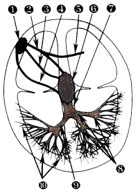
Figure 1. The conducting system of heart.
1 - sinoatrial node; 2 - Bachman internode front canal; 3 - Wenckebach internode average canal; 4 - Thorel internode posterior canal; 5 - interatrial bundle of Bachman;6 - aprioventicular node; 7 - common trunk of Gis node; 8 - left foot of Gis node and its branches;9 - right foot of Gis node; 10 - Purkinje fibers.
4.2. Leads in ECG
Leads can be divided into the single-polar and bipolar ones, as well as leads, which are on the body and inside of it.
ECG method is based on the registration of the potential difference, which arises on the body. Points, between which we register potential difference, are called ECG leads. Potential difference registration is recorded with help of electrodes, which are connected to a galvanometer of electrocardiograph. One electrode is called active or positive, the other is passive or negative. Nowadays the common practice is to use only 12 leads, 6 of which are leads from extremities and 6 from the chest:
- 3 standard leads (from the extremities);
- 3 amplified single-polar leads (from the extremities);
- 6 chest leads.
Four electrodes are used in the extremities leads. Among three of them the potential difference is registered, and the 4th (black) lead is the ground. They have standard color marks and place:
- red (right wrist);
- yellow (left wrist);
- black (right leg);
- green (left leg).
Standard leads. They are the first leads, which were used for ECG registration. They were offered by Einthoven in 1913 (i.e. 10 years after he developed the first ECG galvanometer). These leads are called as standard and marked with numbers I, II, III. Standard leads register such potential difference as:
- lead I - between arms (yellow electrode is positive);
- lead II - between a right arm and a left leg (green electrode is positive);
- lead III - between left extremities (green electrode is positive).
Einthoven represents heart as a point electric current source - dipole. The potential difference registration takes place in frontal plane. Lets imagine that we connect leads, then we get an equilateral triangle, which is named after Einthoven (fig. 4.1 and fig. 4.2). When we trace three axes trough the center of the triangle, we get a three-axial lead system (fig 4.3. A), which makes easier ECG analysis in standard leads. [1]
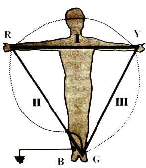
Fig. 4.1. Einthoven standard leads ( R – red electrode; Y – yellow electrode; B –black electrode; G – green electrode).
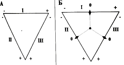
Fig.4.2. A – Einthoven triangle and electrode polarity.
B – Einthoven triangle with perpendiculars to sides.
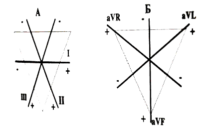
Fig.4.3. Einthoven three-axial bipolar lead system (A) and Golberger three-axial single-polar leads system (B).
Amplified leads from extremities are single-polar leads, used with the same electrodes as for the standard leads. Their location also coincides with standard leads. Active electrode is connected to the positive pole of the galvanometer and located on one of the extremities. The other – combined electrode of Goldberger, which is a combination of the two electrodes from the other two extremities, is a passive electrode.
Marks of these leads are the following:
- a - augmented;
- V - Voltage;
- R - Right ;
- L - Left ;
- F - Foot.
So there are three amplified leads of extremities:
- aVR - amplified lead of right hand;
- aVL - amplified lead of left hand;
- aVF - amplified lead of leg.
We can create a three-axial system of amplified leads of extremities by analogy to a bipolar three-axial system of standard leads (fig. 4.3. B).
4.3. Technique of ECG registration
The technical error, which might occur during ECG registration, can cause serious problems for the further analysis of ECG data. The quality of ECG registration depends on:
- the proper order of electrodes on the chest and extremities;
- the reliability of contact between electrodes and a human body;
- serviceability of electrocardiograph;
- electrocardiograph grounding.
Ability of changing ECG registration parameters is limited by:
- a belt speed during ECG registration;
- choice of the amplitude of register waves and complexes (the amplitude of 1 mV). [7]
5. Block diagram of electronic system
На рисунке 2 представлена структурная схема одноканального кардиографа на базе, которого будет построена система регистрации ЧСС и ЧДД. [9]
Figure two shows a block diagram of a typical single-channel electrocardiograph, which is the base of the electronic system of small test animal HR and FRA registration.
Next blocks used on the block diagram: an amplification block, which is a differential instrumentation amplifier with a set gain factor; an isolation amplifier; a filtration unit, which includes three filters: of low-frequency, of high frequency and a rejecter filter; ADC block and a microcontroller unit.
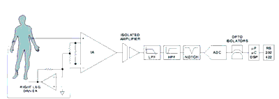
Figure 2. The block diagram of the electronic system.
Figure 2.1. Animation of heart activity registration electronic system. Flash – animation, 1130 frame, 31,8КБ.
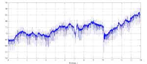
6. Experimental data processing.
To verify our hypothesis, we made an experiment in which we obtained the following data:
a
b
с
Figure.3. Results of a test animal ECG registration.
a - I lead, b - II lead, с - III lead.
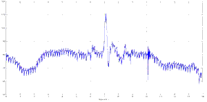
The obtained data were processed using fast Fourier transform (FFT) in the software package MATLAB 7. Results of processing are shown as frequency spectrum in figure 4. [4]
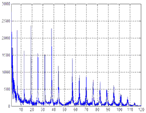
Fig.4. Frequency spectrum
After analyzing the frequency spectrum it is possible to find out several regularities. One of them can be found in the frequency domain of 6.5 Hz and multiples of it. The increase of the power of this frequency is connected with the HR. The other is not so clear, however, after additional analysis it is possible to determine one more frequency domain with increased power, which is nearly 1.4 Hz. Signals of this frequency are connected with the FRA of the test animal. The third regularity implicates the availability of a constant component and low frequencies up to 0.5 Hz. At the high frequency part of the spectrum of initial signal of 50 Hz the disruption of the first regularity, which is connected with the rejecter filter in the electric electrocardiograph with 50 Hz suppression, is observed.
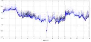
To improve the identification of FRA and HR the initial signal of the electrocardiograph was processed by means of digital filtration. The aim of the filtration is to decrease the influence of the constant component and frequencies up to 0.5Hz. The initial signal was processed with Butterwort digital high-frequency filter with cutoff frequency of 0.5Hz. Whereas such a digital filter has a high lag effect on the oscillogram (figure 5) it is possible to see the zone of transient processes, which lasts about two seconds.
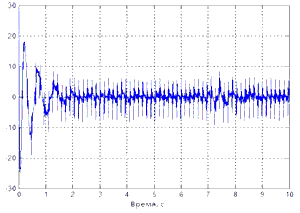
Fig.5. I lead after HF filtration
For further frequency analysis time interval from zero up to two seconds must be removed from the signal, because its presence will lead to additional errors. The frequency spectrum without the part with transient processes (figure 6) is shown on the figure 7.
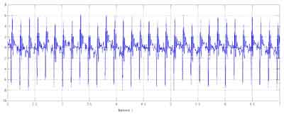
Fig.6. Part of ECG from the 2nd up to the seventh seconds
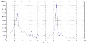
Fig.7. Power spectrum of the ECG part
Analyzing the obtained frequency spectrum it is quite easily to detect frequencies that are connected with HR and FRA. The results of this study shows that using a one channel electrocardiograph and digital processing of data the reliable information as for HR and FRA of test animal can be got. As a result of this research we can say that using the miniature electrocardiograph as an electronic system of small test animal HR and FRA registration is possible and rational, however it requires further digital processing of data.
Conclusion
This study formulates objectives, which are argued by the real necessities of pathophysiologists in a registration system, which are stipulated by improving the effectiveness of medical researches. The miniaturization of the system makes it unnecessary immobilization of the test animal, which will increase the reliability of data. Registration of HR and FRA is based on the analysis of the ECG data, which are obtained with help of the miniature electrocardiograph.
Further studies will allow to build an optimal algorithm for determining HR and FRA from the ECG signal.
References
- Толстой В.А. 22 лекции по ЭКГ: Учебное пособие. – Донецк: ООО «Лебедь», 2000. – 448 с.
- Мурашко В.В., Струтынский А.В. Электрокардиография: Учебное пособие – М.: «МЕДпресс», 1999. – 312 с, ил.
- Рекурсивні цифрові фільтри (розрахунок та синтез структурних схем, розрахунок похибок квантування)/ Геанін В.О., Єрмоленко П.А., Макаренко В.В., Мовчан Т.В., Писаренко Л.Д., Пілінський В.В., Розорінов Г.М., Хотяїнцев С.М. : Навчальний посібник. – Київ: ВПФ УкрІНТЕІ, 2001. – 184с, іл. - Укр.
- Введение в цифровую фильтрацию/Под ред. Р.Богнера и А.Константиндиса. – М.:Мир, 1976. – 364с.
- Голд Б., Рейдер У. Цифровая обработка сигналов. – М.: Советское радио, 1979. – 367с.
- Де Луна А.Б. Руководство по клинической ЭКГ. Пер. с англ. – М.: Медицина, 1993 – 361с.
- Дощин В.Л. Клинический анализ электрокардиограммы. – М.: Медицина, 1982 – 207с.
- Теоретические основы электрокардиологии// под ред. Нельсона К.В. и Гезеловица Д.Б.: пер. с англ. – М.: Медицина, 1979. – 470с.
- Eckart Hartmann. ECG front-end design is simplified with MicroConverter [Электронный ресурс]: – Режим доступа: http://www.analog.com/library
- Amirali Shayan Arani. Noninvasive Cardioneural Signal Extraction. [Электронный ресурс]: – Режим доступа: http://roger.ucsd.edu/Amirali
- Вайсман М.В. Построение алгоритмов и средств испытаний многоканальных цифровых электрокардиографов. [Электронный ресурс]: – Режим доступа: http://ecg.ru/pub
- Antoniadis A., G. Oppenheim. Wavelets and statistics, Lecture Notes in Statistics 103. – Springer Verlag, 1995.
- B. Burke Hubbard. The world according to wavelets. – Wellsley.: AK Peters, 1996.
Note
When writing this abstract the masterwork hadn't been completed yet. The deadline of completion is December 1, 2010. Full text of the work and materials on the subject-matter can be obtained from the author or her scientific adviser after that date.
N.B.:
DonNTU *
Master's portal *
Biography * Abstract
Abstract