
Abstract
Content
- Introduction
- 1. Goal and tasks of the research
- 2. Description of the current methods of classification of image processing
- 2.1 Methods of comparison standards
- 2.2 Methods based on the geometric characteristics
- 2.3 Methods of optical flow
- 3. Structure of the masters
- 3.1 Selecting an object of diagnostics
- 3.2 Choice of methods for the classification of image processing. Markov models
- 3.3 Structure of SCS
- Conclusion
- References
Introduction
Telemedicine – the use of a telecommunications technology advances in health care. Telemedicine can improve the effectiveness of treatment and diagnosis to a new level. With the help of telemedicine technology can, for example, a remote patient, to provide high-quality medical care. Doctors can make a diagnosis on the basis of real-time or via e-mail images of X-rays, computer tomograms, etc. Due to the fact that often the bulk of narrow specialists in various fields of medicine works in specialized medical centers in large cities, this has led to a certain centralization of medical care. Advances in telemedicine eliminates the need for physical presence of a specialist on the spot.
Telemedicine – a direction to the intersection of several fields – medicine, telecommunications, information technology, and education. The direction of this new, but rather demanded recently.
This SСS can quickly and efficiently handle the large number of images and a preliminary diagnosis. Then the results are transmitted to specialists, who in turn decide on further treatment of the patient.
1. Goal and tasks of the research
The aim is to study and selection of image processing techniques for bone SCS. The development of SCS diagnosis of fractures.
The main objectives are:
1. Analysis of existing computer systems diagnosis of fractures.
2. Research and selection of image processing methods for classifying fractures for the SCS.
3. Experimental study of selected methods and adapting them to SCS.
4. Development of the structure of SCS and MS, PS, IS to SCS.
2. Description of the current methods of classification of image processing
2.1 Methods of comparison standards
Comparison of the standards is to allocate the necessary areas of the image, and then comparing these areas for two different images. Each region increases coincided measure of similarity of images. This is also one of the historically first image recognition methods. For comparison, areas used simple algorithms, such as per-pixel comparison.
The disadvantage of this method is that it requires a lot of resources like storage areas, and to compare them. In view of the fact that used a simple matching algorithm, the image should be removed in a strictly prescribed conditions: not allowed significant changes angles, lighting, etc.
2.2 Methods based on the geometric characteristics
One of the earliest methods – an analysis of the geometrical characteristics. Originally used in criminology and has been developed in detail there. Then came the computer implementation of this method. Its essence lies in the allocation of a set of key points (or areas) and then allocating a set of attributes. Each feature is either the distance between key points, or the ratio of these distances. Unlike the comparison of elastic graph distance is not chosen as the arc graphs. Sets of the most informative features are distinguished experimentally. In recognition of symptoms compared to the unknown, for example, a person with symptoms that are stored in the database. The problem of finding the key points close to the complexity of directly recognizing and finding the correct key points on the image largely determines the success of recognition. Therefore, the image should be no interference, hindering the process of finding key points. Lighting is desirable uniform and equal for all images. In addition, must have a front perspective, perhaps with minor variations.
Thus, this method imposes stringent requirements on the shooting conditions, needs a reliable mechanism for finding the key points for the general case. It also requires the use of more sophisticated classification methods, or construct a model of change.
In general, this method is not the best, but for some specific tasks perspective. These tasks include the control of Document, when you want to compare the face image obtained in the moment with a photograph in the document. At the same time other images of this person is not available and therefore mechanisms of classification, based on an analysis of the training set are not available.
2.3 Methods of optical flow
Optical flow algorithms are mainly used for the analysis of movement. Using two or more successive image frame, we can calculate the two-dimensional vector field, called optical flow, which reflects the actual or most probable displacement of image points from frame to frame.
Optical flow is calculated for any two images measures for compliance with pictures. These two images are considered consecutive frames. Then we calculated the vector field best showing one image to another, in the sense of minimize the distance between the images and taking into account the geometrical restrictions, such as the relative positions of image points.
The algorithm finds the most appropriate units. The search is performed hierarchically, starting with the big blocks, and then breaking them into smaller blocks. Thus, the pyramid was built matching images.
Using the vector field of optical flow, built the various measures of conformity, by which the base was an image that is closest to the unknown.
The disadvantages of this method is especially true of its computational complexity. On a dedicated parallel computer to compare two images took more than a minute. The method is unable to extract a compact feature set for the storage and retrieval in the database.
3. Structure of the masters
3.1 Selecting an object of diagnostics
The object of this study is the image of the femur (radiographs).
Femur – is the largest, longest bone in the human skeleton. Both ends of the femur are involved in the formation of large joints – the hip and knee. For anatomical localization of fractures are: the proximal end of the body, the distal end. In this paper we consider the body fractures of the femur (diaphysis).
Fractures of the shaft divided into simple – it is a simple violation of the integrity of the diaphysis of the circular, wedge – fracture of one or more intermediate fragments, which persists after repositioning the main contact between the fragments, as well as complex – a fracture of one or more intermediate fragments, which, after repositioning there is no contact between the main fragments. Based on the description, the following characteristics to determine the type of fracture (Table 1):
Table 1 – The characteristics of the type of fracture
| Simple | Wedge | Complex | |
| Number of Parts | 2 | >2 | >2 |
| The shape and size of units | 2 large pieces that can be combined | 2 major parts, the rest of all shapes and sizes. Perhaps the visual alignment of the fragments. | 2 major parts, the rest of all shapes and sizes. Visual alignment of the fragments can not be. |
In turn, there are simple fractures: spiral, oblique and transverse. Wedge fractures are divided into spiral, bending wedge and fragmented. Complex fractures: spiral, segmental, and irregular.
")
Picture 1 – Types of hip fractures
(animation: 5 frames, 5 cycles of repeating, 115 kilobytes)
3.2 Choice of methods for the classification of image processing. Markov models
Selected basic techniques that will be used in image processing in the SCS. Methods based on Markov models. Markov models are a powerful tool for modeling various processes and pattern recognition. By their nature, Markov models allow for the direct space-time characteristics of signals, and therefore are widely used in speech recognition, and recently – pictures.
Digital image – the essence of a random two-dimensional digital signal which is observed by the system. The sequence of observations О=о1, о2, …, оT, where оt – signals are sorted by the spatial relations can be extracted from the image in various ways. For this reason, the descriptive ability of models obtained may vary.
Each model λ=(A, B, π) is a set of N states S={S1, S2,…, SN}, between which transitions are possible. At any time the system is in a very specific condition. In the transition to each state is generated by the observed symbol that corresponds to the physical signal from the output of the simulated system. The character set for each state V={v1, v2,…, vM}, the number of characters M. There are also models in which the character set for all states the same.
The symbol in the state qt=Sj at time t is generated with probability:
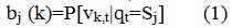
The set of all these probabilities is a matrix B={bj(k)}, where bj(k) – the probability of the k-th parameter in the j-th state.
The matrix A={aij} determines the probability of transition from one state to another state:

It is believed that A does not depend on time.
The same model has a likelihood of initial states π=πi, where πi=P[q1=Si] – the probability that at the initial time the system will be in the i-th state.
The model λ=(A, B, π) to the set parameters can be employed to generate a sequence of observations. To do this, by chance, in accordance with the initial probabilities π the initial state is chosen, then at each step the probability of B is used to generate the observed characters, and the probability of A – to select the next state. The probability P to generate a model of the sequence of states λ:
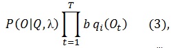
where Q=q1, q2,…,qT – the sequence of states.
It is assumed that the observations are statistically independent.
In pattern recognition hidden Markov models are applied as follows. Each class i has its own model of λ. Recognizable image is represented as a sequence of observations O. Then, for each model λ is computed the probability that this sequence could be generated by this particular model. Modelλj, received the highest probability is considered to be the most appropriate, and the image belongs to class j.
3.3 Structure of SCS
The basic sequence of operations, automatic classification shown on picture 2:

Picture 2 – Automatic classification
In most cases, the automatic classification is performed in three stages:
1. Stage of training images include the direct input of images from the camera and pre-processing. Pre-treatment consisting in extreme close study to the reference image and normalized. Most often for medical images is spatially invariant operation, shift, change brightness, change contrast, quantization and geometric transformations (scaling, rotation axis). The theory of these transformations are well developed and usually does not cause difficulties in the use of modern computers.
2. Feature extraction, in which the function representing the processed image undergoes functional transformation, identifies a number of the most essential features, which are encoded by real numbers. Feature extraction is the mathematical transformation of the image depending on the task analysis. This may be a subtraction from a reference, subtract a constant component to avoid disturbing shadows, differentiation, or to highlight the contour of the autocorrelation, frequency filtering, and many others. The correct choice of processing algorithm is crucial for the next phase of transformation and poses the greatest difficulty.
3. Classification of signs. The resulting sets of the previous operation of real numbers, describing the selected features, compared with the reference numbers laid down in the machine memory. Computers on the basis of this comparison classifies the image, assigns it to one of the known species, such as the norm or pathology.
Conclusion
In the field of modern information technology focuses on improving the quality of radiographs for improved work with them, also made all kinds of automated processing of medical images in order to better define the parameters and characteristics.
Selected an object of diagnosis, study materials on diagnosis of fractures, fracture classification, analyzed the existing methods of image processing, their advantages and disadvantages of selected optimal methods of image processing for the SCS.
References
- Телемедицина в России [Electronic resource]. – Аccess mode: http://www.cplire.ru/mac/telemed/index.htm .
- Кулешов, С. В. Применение СММ для распознавания лиц [Electronic resource]. – Аccess mode: http://exos.org.ru/download/frbook.pdf .
- Компьютерная графика и мультимедиа [Electronic resource]. – Аccess mode: http://cgm.computergraphics.ru/ .
- Перелом проксимального отдела бедренной кости [Electronic resource]. – Аccess mode: http://www.eurolab.ua/diseases/1456/ .
- Учебно-методическое пособие [Electronic resource]. – Аccess mode: http://www.argo-shop.com.ua/article-4366.html.
- Перелом бедренной кости [Electronic resource]. – Аccess mode: http://www.vrach-travmatolog.ru/perelom-bedrennoy-kosti.htm.
- Перелом костей [Electronic resource]. – Аccess mode: http://www.vrach-travmatolog.ru/perelomy-kostey.htm.
- Моделирование. Вероятностные методы диагностики [Electronic resource]. – Аccess mode: http://www.omsk-osma.ru/img_pulpit/mbf/lec_21.Modelirovanie.pdf.
- Сектор лучевой диагностики и медицинской визуализации [Electronic resource]. – Аccess mode: http://www.arcerm.ru/clinicldiag.html.
- Медицинские изображения и их обработка [Electronic resource]. – Аccess mode: http://gridclub.ru/library/publication....
- Стоматологический блог врача-стоматолога [Electronic resource]. – Аccess mode: http://stomklin.ru....
- Распознавание изображений [Electronic resource]. – Аccess mode: http://dima78.livejournal.com/42168.html.
- Цифровые рентгенографические системы [Electronic resource]. – Аccess mode: http://stomfak.ru/rentgenodiagnostika-v-stomatologii....