
Abstract
Contents
- Introduction
- 1. Actuality of the theme
- 2. Purpose
- 3. Main tasks
- 4. The mathematical methods used in the work
- 4.1 Filtration
- 4.2 Contrast and binarization
- 4.3 Allocation of the contours of objects
- Conclusion
- References
Introduction
In recent years, the introduction of computers in various areas of human life is increasingly. Medicine is no exception. Modern ultrasonography machines are equipped with a variety of additional programs and scan modes that open up new perspectives in the diagnosis of various diseases. Analysis of the images obtained with the ultrasonography (the ultrasound images), and the subsequent programing analysis, gave the most complete information about the states of the liver, as well as the presence of pathologies.In the presence of modifications may determine the degree of homogeneity, location, size and area.
1. Actuality of the theme
The liver is often called the laboratory of the human body.In this organ happens about 20 million chemical reactions per minute.The problem of maintaining the health of the liver today more than ever.Almost half of the population are observed certain changes in the liver.Recovery the liver - not only very difficult but also very expensive.
To to assist with diagnosing doctors, as well as for earlier diagnosis requires a computer system diagnostics with special software that will simplify the process of finding tumors and to determine the necessary parameters. This will provide the doctor to more accurately estimate the size of tumors, accelerate the healing process.
2. Purpose
Development of a computer system processing of ultrasonography images of liver with the a view to finding tumors, identifying the necessary parameters that will help to more accurately assess the current clinical situation.At the stage of invasive methods, such as biopsies. And thus, reduce the risk of complications.
3. Main tasks
main tasks that is necessary to solve in the development a computer system diagnostic pathology of the liver:
- Determination methods and parameters for preprocessing of images to improve their quality.
- Selection of features that characterize the state of the tumor and allow us to solve the problem of automatic classification of pathologies.
- The selection of methods and software development to obtain the necessary evidence.
- The selection of methods the classification of identified pathologies produced by signs, as well as the formulation of a preliminary diagnosis.
Object of research: the images of the liver, obtained by the ultrasonography machine.
Subject of research: methods of analysis ultrasound images of the liver, allowing the correct preliminary diagnosis.
4. The mathematical methods used in the work
The simplest form of digital image processing is f(n1,n2), 1 <= m1 <= N1, 1<= m2 <= N2. perform the same functional converting each element of the matrix, regardless of its position and the value of other (adjacent) cells.
This processing is called elementwise image conversion. It takes the value of each element f in the new value of g according to a predetermined functional relationship (1):
| g = g(f ) | (1) |
4.1 Filtration
The best results for the conservation of differences of shades, various borders and local brightness peak incorrupted by impulse noise image may be achieved by the use of the median filter.
The median filter is a window W,sliding over the image field, covering the an odd number of samples.the central countingIs replaced by the median of all elements of the image, caught in the window.The two-dimensional median filter with a window W is defined by the formula (2):
 | (2) |
where y(n1,n2) — luminance value of the original image pixel with coordinates n1 and n2; х(n1,n2) — luminance value of the filtered image pixel with coordinates n1 and n2.
4.2 Contrast and binarization
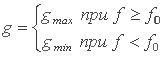
One of the basic and easiest ways - is to build segmentation using a threshold of brightness.
Operation threshold separation (3) consists in comparing the brightness of each pixel with the given threshold value f0.
 | (3) |
where f0 - a "threshold" value of brightness. When the threshold processing main question is to choose a threshold f0.
4.3 Allocation of the contours of objects
After application of the methods described above performedoperation of the image edge detection.In particular, for detecting usethe method Roberts (4):
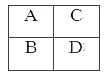 |
Picture 1 – Aperture use the method Roberts
 | (4) |
where – A' - the new value of the brightness which is necessary to reassign the point A, the initial aperture.Aperture is necessary to move on the image left - right, top - down.
Conclusion
After the application of all the methods described in the work, and, in appropriate form coefficients we obtain the result (pict.2).
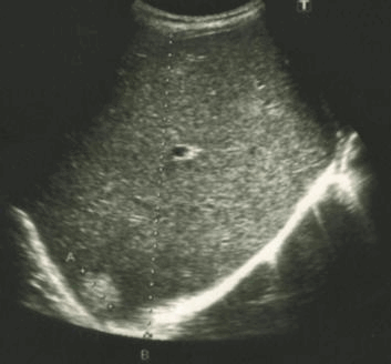
Picture 2 - Result of work
In my work I tried to choose the most appropriate methods contrasting and filtration out those that are used in existing processing systems ultrasonographyimages. Using the methods of binarization and contour, I select the objects in the image.
This master's work is not completed yet. Final completion: December 2013. The full text of the work and materials on the topic can be obtained from the author or his head after this date.
References
- В.В. Митькова, Клиническое руководство по ультразвуковой диагностике. Том 1 //под ред. Митькова В.В. - М.: Видар, 1996
- К.П. Майер Гепатит и последствия гепатита: практическое руководство. - М., 2000
- Миллер Р. Теория переключательных схем / Р. Миллер. – М.: Наука, 1971. – Том 2: Последовательностные схемы и машины. – 304 с.
- В.А. Сойфер Компьютерная обработка изображений. Часть 1. Математические модели // Соросовский образовательный журнал, 1996, №2
- В.А. Сойфер Компьютерная обработка изображений. Часть 2. Методы и алгоритмы // Соросовский образовательный журнал, 1996, №3
- У.Л. Прэтт Цифровая обработка изображений. Кн.2. - М.: Мир, 1982
- Hamarneh G., Gustavsson T. Combining Snakes and Active Shape modles for Segmenting the Human Left Ventricle in Echocardiographic Images. IEEE Com-puters in Cardiology 2000; vol 27
- Hiransakolwong N., Windyga P.S., Kien A. Hua FASU: A Full Automatic Segmenting System for Ultrasound Images. School of Electrical Engineering and Computer Science University of Central Florida 1998