
Abstract
Content
- 1. Relevance
- 2. Research goals and objectives, expected results
- 3. Preparation of the vascular bed
- 3.1. Binarization of image
- 3.2. Skeletezation of image
- Conclusion
- References
1. Relevance
Heart disease (heart disease) – disruption of normal functioning of the heart. Includes defeat epicardium, pericardium, myocardium, endocardium, valvular, heart vessels.
Heart disease can occur a long time in a latent form, clinically does not prove itself. Along with various tumors, these diseases are now the major cause of premature deaths in developed countries.
Uninterrupted operation of the circulatory system, consisting of the heart as a muscular pump and a network of blood vessels - a necessary condition for the proper functioning of the body.
Heart and vascular disease today represent a real threat to humanity. And it is - no exaggeration. They are a major cause of death and disability, even beating cancer. If only a few decades ago, heart disease was mainly lot of older people, but now there is their "rejuvenation" - more often they arise under the age of 40 years.
Treatment of cardiovascular diseases is a very complex issue, which needs special expensive medical equipment and supplies, highly skilled, and most importantly - the availability of care for the population. Unfortunately, only in the advanced economies, this help is available and effective - in Western Europe, the U.S. and Israel. Is it possible prevention of cardiovascular disease? Doctors all over the world working on this problem, say that prevention is not only possible but also necessary.
It is possible to diagnose the early stages of cardiovascular disease based on analysis of human microvascular.
2. Research goals and objectives, expected results
Ph.D. Konstantinova and Ivanova developed a method of quantify images in the diagnosis of bulbar conjunctiva microcirculation in cardiovascular pathology [1].
This method involves the calculation of several groups of coefficients, which are calculated based on the relative positions, space, size and type of vessels in the image area of ??the bulbar conjunctiva 1 mm2. Many calculations in this method involves representation of the image in the form of a skeleton of the vascular bed.
Goal of this work is the development of the evaluation system of the cardiovascular system to increase the speed and accuracy of diagnosis by doctor cardiologist by automatically calculating the coefficients, which allows the doctor to spend more time analyzing them.
Main objective is the isolation of the vascular bed from an existing image and building a model of the skeleton of this channel, on which in consequence will be calculated coefficients state microvasculature and provided in conjunction with the standard values??. The totality of these coefficients will help a doctor to draw conclusions and decide on the state of the cardiovascular system of the patient.
Since estimated geometric characteristics of vessels, it is logical to assume that as the initial data required to submit an image as a skeleton. To get an idea of the vascular bed in such a database, I need to:
- Binarize an image. At this stage, the source image is converted into binary: pixels belonging vessels stained in the object color and the background color in the rest. Conditionally object color set to black color, and background color – white.
- Skeletonize a binary image. This phase involves the construction of a curve equidistant from the borders of the vessels.
Object of research: the process of diagnosis of the cardiovascular system.
Subject of research: dimensioning and analysis of proper diagnosis of these parameters.
Within the master's work is planned to obtain relevant scientific results in the following areas:
- Assessment of the cardiovascular system in the image of bulbar conjunctiva obtained without the help of specialized instruments.
- Definition scope of the method [1].
- Modification of known methods of image analysis to improve accuracy and reduce the analysis time.
For experimental evaluation of the theoretical results and the formation of the foundation of further research, as practical results is planned to develop a system of analysis of the cardiovascular system of the bulbar conjunctiva in the image with the following properties:
- Having base of normative values of ??computed parameters.
- Unified approach to image analysis.
- Generation of reportabout calculated parameters in comparison with their normative interval.
Preparation of the vascular bed
Binarization of image
There are many methods of image binarization that may generally be represented as a method of segmentation or clustering methods (with the number of clusters equal to two ). Any binarization is performed on the halftone image with a color depth of 8 bits per pixel, so the original image is converted to grayscale in advance.
There are two areas of concern in the process of binarization: correct definition of object pixel background pixels, binarization time.
Conventional methods provide a good indicator in terms of time, but the resulting image is very "noisy", which ultimately may affect in the future, the construction of the skeleton. Results of methods based on genetic algorithms has better quality, but they work slowly and heavily dependent on computer processing power.
The optimality criterion was used condition "Leave as much information about the vessels, with the least amount of noise". Various methods of normal binarization have been considered and is best approached under this criterion method is Sauvola, which is a method based on local thresholds:
where pixel – current pixel, mean – average color value in the image, k – sensitivity, r – radius of the window, stdev – standard deviation.
Average color value is calculated by the following formula:
where n – image height, m – image width, pixelij – color value of a pixel.
The standard deviation is calculated using the following formula:
This method was selected because image area is constant, so in the process of selection size of a window and other factors for the method, we can obtain better results than other similar methods. Coefficients used for the selection of genetic algorithm working with real numbers. Among the various options for crossover and mutation were tested 10 combinations. Result of work represented by the following table:
| Mutation | |||
|---|---|---|---|
| Uneven | Random | ||
| Crossing | Arithmetic uneven | 5123 | 4192 |
| Arithmetic even | 3264 | 4218 | |
| Flat | 4565 | 3924 | |
| Min-max | 3358 | 3065 | |
| Simple | 4615 | 3788 | |
Fitness function is the condition of maximum similarity of the result and decire. Numerical representation was the number of pixels that do not coincide with the result, which was determined by an expert:
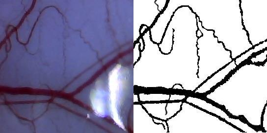
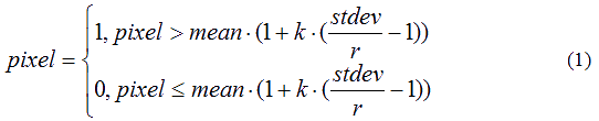
Figure 1. Original image (left) and the desired result (right)
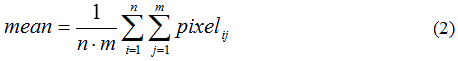
Process of method works with min-max crossover and random mutation is shown in Figure 2.
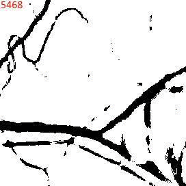
Figure 2. Process of achieving the desired result with the min-max crossover and random mutation
(animation: 36 frames, fixated, 636 kilobytes)
As a result, the optimal values ??of the coefficients for the Sauvola method are Size = 15; k = 0,051; r = 503. This method for given values ??as precisely as possible creates a binary image.
However, due to the light area of ??the image, resulting in noise appears. In order to reduce the noise level of binarization methods was chosen such that most accurately be defined light areas. Such was the method Benrsen, which works according to the following formula:
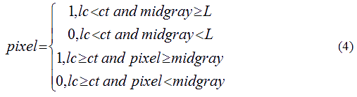
where lc – local contrast value, ct – threshold of contrast, midgray – the average value of the gray.
L calculated from the formula:

where p – color depth. Since being processed halftone image with a color depth of 8 bits, in this case L=28-1=128.
Similarly, as for the method Sauvola, was picked up by crossover and mutation method:
| Mutation | |||
|---|---|---|---|
| Uneven | Random | ||
| Crossing | Arithmetic uneven | 1402 | 1597 |
| Arithmetic even | 1110 | 663 | |
| Flat | 1773 | 678 | |
| Min-max | 1256 | 1090 | |
| Simple | 1369 | 1836 | |

Figure 3. Expected (left) and resulting (right) result
Optimal values ??of the coefficients for the Benrsen method are Size = 7, ct = 76, midgray = 132.
Applying as a mask for the first image obtained result of method Benrsen, we obtain the final result:
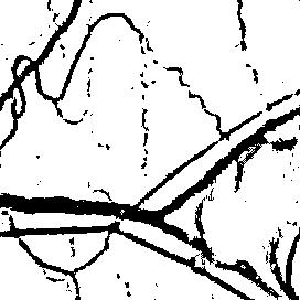
Figure 4. Binary image
3.2. Skeletezation of image
Skeletonization process is that the blood vessels necessary for producing a binary image represented in a logical structure.
There are several methods of skeletonization, each of which has a series of positive and negative factors.
The method of wave spreading [11] is the simplest method skeletization as for his work there is no need to reduce the thickness of the vessel to 1-wide pixel, but you must specify the starting point from which this method will "spread a wave". Positive factor is the speed, which does not require frequent reference to the image. The downside of this method is that the result will be detected only one closed vessel, which is unacceptable in the current work.
Zong-Suan's method [12] based on the templates of deleting pixels that do not belong to the skeleton. This algorithm is performed as long as the result is not removed any pixel that assumes a multiple pass image, but the result is the skeleton of the vascular bed wide 1 pixel, as required. Further transformation of the logical structure of the resulting skeleton is over.
Template method is an extension of Zong-Suan's method, complementing its algorithm own templates whereby the pixels will be deleted. Solution of the patterns shown in Figure 5. As you can see, the result was obtained by the skeleton of the vascular bed with "noise". To eliminate it is necessary to conduct research and add to the list of templates, which would allow to significantly improve the quality of the process.
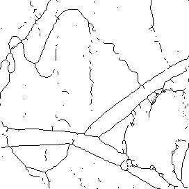
Figure 5. Skeleton of an image
Conclusion
The paper considers the importance of the problem of cardiovascular disease diagnosis by images of bulbar conjunctiva, on the basis of the methodology [1]. Shown that its application is required to solve such problems as digital image processing and binaryzation and skeletonization. The analysis of iterative and evolutionary methods binaryzation and skeletonization, to identify the best solution at the moment binaryzation (Sauvola and Benrsen) and skeletonization (templates method). For image binaryzation experimentally determined values ??of the parameters of the Sauvola method. However, found that as a result contains a lot of noise, which is eliminated by postprocessing. Experimental analysis has shown that this method is expedient to use Benrsen, which gave acceptable results. For each method using a genetic algorithm was chosen the best options.
Consider the solution of the problem of obtaining topological skeleton obtained binary image. It is shown that good results are obtained by applying the templates method. Thus, the obtained results allow us to further perform the calculation coefficient values ??necessary for decision making.
Direction of future work is to create a system for automatic calculation of the parameters of the cardiovascular system based on the methods discussed above.
References
- Е.Э. Константинова, Л.А. Иванова, Метод количественной оценки изображений бульбарной конъюнктивы в диагностике состояния микроциркуляции при сердечно сосудистой патологии, Беларусь, 2004 г., 10 с.
- Wells R., Goldstone J. Rheology of the cell and capillary flow.– In: Rheology of biological systems.— Gabeknik, 1973.– P. 186.
- Yvonne-Tee G.B., Ghulam Rasool A.H., Sukari Halim A. et al. Noninvasive assessment of cutaneous vascular function in vivo using capillaroscopy, plethysmography and laser-Doppler instruments: Its strengths and weaknesses. // Clinical Hemorheology and Microcirculation, 2006.– N 34 (2006).– P. 473–457.
- Maggio E. Microhaemocirculation: observable variables and their biological control. Springfield, Thomas, 1965.– 194 p.
- Bloch E. Microscopic observation of the circulation blood in the bulbar conjunctiva in man in health and disease // Ergebn. Anat. Entwikl.,– Gesch., 1956.– N 35.– P. 1–98.
- Knisely M.H. Intravascular erythrocyte aggregation (blood sludge) // Handb. of physiology, Sect. 2: Circulation.– London, 1965.– Vol. III.– P. 2249–2292.
- Чернух А.М., Александров П.Н., Алексеев О.В. Микроциркуляция.– М.: Медицина, 1984.– С. 421–447.
- Орлова А.Н. Вклад А.И. Нестерова в учение о микроциркуляции (к 85-летию со дня рождения) // Вопр. ревматизма, 1981.– № 1.– С. 58–61.
- Люсов В.А., Белоусов Ю.Б. Гемостаз и микроциркуляция при сердечно-сосудистых заболеваниях // Терапевт. архив, 1980.– Т. 52.– С. 5–14.
- Богоявленский В.Ф. Значение биомикроскопии сосудов конъюнктивы для диагностики прогрессирующего атеросклероза // Казанский медицинский журнал, 1969.– № 3.– С. 65–70.
- Применение волнового алгоритма для нахождения скелета растрового изображения [Электронный ресурс] // Режим доступа: http://ocrai.narod.ru/vectory.html
- Weian Deng, S. Sitharama Iyengar and Nathan E. Brener, A Fast Parallel Thinning Algorithm for the Binary Image Skeletonization, International Journal of High Performance Computing Applications [Электронный ресурс] // Режим доступа:http://citeseerx.ist.psu.edu/viewdoc/download?doi=10.1.1.226.2059&rep=rep1&type=pdf