
Abstract
Content
- Introduction
- 1. Relevance of the topic
- 2. The purpose and objectives of the study, the planned results
- 3. Analysis of the control object
- 3.1 Blood parameters and characteristics
- 3.2 Methods for measuring blood oxygen saturation
- 3.3 Criticism of methods for measuring blood parameters
- 4. Technical requirements for development
- 5. Development of the structural scheme
- 6. Evaluation of metrological parameters
- 7. Development of the device design
- Conclusions:
- List of sources
Introduction
Saturation is the saturation of a liquid with gases. In medicine, saturation is the concentration of oxygen in the blood, which is expressed as a percentage.
Low oxygen saturation of the blood leads to a weakening of the cardiovascular and immune system, the brain slows down. Subsequently, not only does the physical condition weaken, but mental retardation is also observed.
95-100% is considered the norm of arterial blood saturation. At 94% hypoxia develops and measures are required to prevent it, less than 90% - the situation is critical, the patient needs emergency medical care.
At the present stage of development of science, there are several methods for determining the degree of blood oxygen saturation: invasive, non-invasive, measurements in real time and laboratory methods.
Given the need for accurate and rapid determination of oxygen saturation in human blood, there is a need for a device capable of meeting these requirements.
1 Relevance of the topic
Non-invasive pulse oximetry has some drawbacks [3], including the change in work in bright light, moving objects, the presence of dyes (nail polish), the need for accurate positioning of sensors. Errors in the readings can be associated with improper imposition of the device, shock, hypovolemia in a patient when the device cannot catch the pulse wave. Carbon monoxide poisoning can even show 100% saturation, while hemoglobin is saturated not with oxygen, but CO.
The master's work is devoted to the urgent task of developing a device for invasively measuring the saturation of a person’s arterial blood with oxygen.
2 The purpose and objectives of the study, the expected results
The purpose of the work is to develop an electronic device for invasively measuring the content of oxyhemoglobin in human arterial blood.
Tasks to be solved to achieve the goal:
- Choose a mathematical model of the formation of the analytical signal by the invasive method;
- Set the conversion characteristic of the measuring transducer of the reflection coefficient (partial pressure) into the value of the photocurrent of the radiation detector.
- Build a mathematical model of the transformation of partial pressure - saturation.
- Build a model of a measuring instrument for estimating metrological parameters.
- Develop a block diagram of the device. Offer circuit solutions for structural components.
- To develop the structural elements of the device for the invasive measurement of the content of oxyhemoglobin in human arterial blood.
3 Analysis of the control object
3.1 Blood parameters and characteristics
Blood is a liquid tissue circulating through the vessels, transporting various substances within the body and providing the nourishment and metabolism of all the cells in the body. The red color of blood gives hemoglobin contained in red blood cells.
Hemoglobin is the main component of red blood cells and provides:
1) the respiratory function of the blood due to the transfer of O2 from the lungs to the tissues and CO2 from the cells to the lungs;
2) regulation of the active reaction (pH) of the blood, possessing the properties of weak acids (75% of the buffer capacity of the blood).
Normally, hemoglobin is contained in the blood in the form of three physiological compounds:
1) oxyhemoglobin (HbO2) - hemoglobin, which attached O2; is in arterial blood, giving it a bright scarlet color;
2) restored, or reduced, hemoglobin, deoxyge-moglobin (Hb) - oxyhemoglobin, donated O2; located in venous blood, which has a darker color than arterial;
3) carbhemoglobin (HbCO2) - the combination of hemoglobin with carbon dioxide; contained in venous blood.
Hemoglobin can also form pathological compounds:
1) Carboxyhemoglobin (HbCO) - connection of hemoglobin with carbon monoxide (carbon monoxide);
2) Methemoglobin (MetHb) is a compound in which, under the influence of strong oxidizing agents (aniline, bertolet salt, phenacetin, etc.), heme iron is converted from ferric into trivalent. When a large amount of methemoglobin accumulates in the blood, the transport of oxygen to the tissues is disturbed, and death can occur.
3.2 Methods for measuring blood oxygen saturation
One of the main indicators of a normally functioning organism is the saturation of arterial blood with oxygen.
Oxygen saturation is the ratio of the amount of oxyhemoglobin to the total amount of hemoglobin in the blood, expressed as a percentage. Saturation is denoted by the symbols SaO2 or SpO2.
This parameter is reflected in the number of red blood cells. Methods for determining it consider below.
Pulse Oximetry
Depending on how saturated hemoglobin is with oxygen, the length of the light wave that it can absorb is changing. This principle is based on the action of a pulse oximeter consisting of a light source, sensors, a detector and an analyzing processor.
The light source emits waves in the red and infrared spectrum, and the blood absorbs them, depending on the number of oxygen molecules bound by hemoglobin. Associated hemoglobin captures the infrared stream, and non-oxygenated - red. Unabsorbed light is recorded by the detector, the device calculates the saturation and outputs the result to the monitor. The method is non-invasive, painless, and it takes only 10-20 seconds to complete.
Today, two methods of pulse oximetry are used: transmission and reflected.
Direct blood spectrophotometry
Direct blood spectrophotometry is used in fiber optic oximeters used to evaluate the oxygenation of venous blood (Sv02). For this purpose, special catheters of the subclavian vein or pulmonary artery are used, usually used to determine the parameters of intracardiac hemodynamics and additionally containing two isolated optical fibers.
The working ends of the fibers with optical nozzles are fixed to the lateral catheter placed in the vessel under study. The opposite ends of the fibers are connected to an optoelectronic converter. The input of the "transmitting" fiber is connected to the source of the probing radiation, the input of the receiving
is connected to the photodetector of the spectrophotometer. Thus, on the output of the photodetector, a signal is formed that is proportional to the fraction of light scattered from a certain volume of blood surrounding the tip of the catheter in the vessel. Measurements are carried out at three wavelengths of 800, 700, 670 nm, which improves the accuracy of determining Sv02.
Blood oxygen tension monitoring
In clinical practice, direct and percutaneous methods of determining the oxygen pressure in the blood are used. In the direct assessment of oxygen tension in arterial blood, blood sample analysis is used. For this purpose, the Clarke oxygen electrode is used, which is an electrolytic cell separated from the test blood by an oxygen-permeable membrane.
For the percutaneous method of determining P02 used in monitor instruments, membrane sensors are used that contain a Clark electrode and a heating element. The electrode membrane is brought into contact with the skin, which is heated to a temperature of about 44 ° C. Under the action of heating, oxygen from capillary vessels diffuses into the epidermis, and then into the electrolytic cell, where the measurement takes place.
3.3 Criticism of methods for measuring blood parameters
Blood oxygen tension monitoring
Errors in the determination of Ptc02 values depend on skin thickness, subcutaneous blood flow, physiological factors affecting the delivery of O2 to the skin surface (decrease in cardiac output, blood pressure, appearance of central vasoconstriction).
Sources of error at pulse oximetry
Dyes injected into the blood, affect the readings of pulse oximeters.
Errors in determining the patient's condition according to SpO2 may occur due to the masking of the decrease in the PO2 value, which may occur before a significant drop in SpO2 begins. This circumstance is explained by the course of the HbO2 dissociation curves.
Errors can occur with low tissue perfusion or severe vasoconstriction due to weak pulsation at the location of the sensor device. It should be noted that in case of severe hemodilution, anemia, and blood loss, high SpO2 levels do not guarantee a safe level of oxygen delivery to the tissues, since the total oxygen capacity of the blood may be insufficient.
Considering the advantages and disadvantages of the methods described, it was decided to develop an electronic device for invasively measuring the content of oxyhemoglobin in human arterial blood.
4 Technical requirements for development
- analyzed component: Sa;
- measurement range of saturation,%: 75-97;
- error,%: ±2;
- rated supply voltage, V: 220;
- nominal frequency of the supply voltage, Hz: 50;
- range of supply voltage deviation from the nominal value,%: from +10 to -10.
5 Development of the structural scheme
To implement a measurement device, the measuring transducer must form an optical flow and deliver it to the analyte. The radiation parameters must be such that the interaction with the analyte leads to a change in both the spectral composition and the magnitude of the radiation flux. To implement all these actions, it is necessary to ensure the spectral composition of the radiation flux, which coincides with the characteristic parts of the reflection spectrum of the analyzed medium.
According to the data of [1], the formation of an analytical signal is most efficiently performed by the two-frequency method. One range of wavelengths should correspond to the most sensitive region, where the change in the spectral composition and flux has the greatest value. The second range should correspond to the region with the minimum parameters and perform the functions of the formation of the comparative signal.
At wavelengths of 625 nm and 810 nm, optical signals are formed using two light-emitting diodes. As a means of delivery of optical radiation and the reflected signal is recommended to use optical fiber [1]. The optical signal is introduced into the optical system, delivered to the end of the fiber, then the fiber is delivered to the analyzed medium, where it is reflected, refracted and then re-enters the fiber. Moving, propagating, in the opposite direction, the optical signal enters the photosensitive element (photodiode), from the output of which the photocurrent value is taken.
In the process of transmitting optical signals according to the optical scheme (Fig. 1) (it consists of: an objective lens with which the radiation flux from the radiation source is formed as a narrow flux and introduced into the fiber, the fiber itself itself, splitter, photodiode) changes in flux is carried out not only due to reflection from the test object, but also due to the loss of optical radiation at the interfaces between media with different optical density. These boundaries are: air is the objective lens, the lens is air, air is the end of the light guide. The transmission coefficient of the interface is defined as:
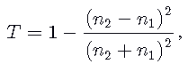
where n1 and n2 are the refractive indices of adjacent media.
A unidirectional splitter allows streams directed from the LED into the light guide of the catheter to be transmitted almost without loss. The reflected streams from the blood into the light guide splitter divides into two equal parts. Half of the signal flow is transmitted to the photodetector. The second half goes to the emitter, where it is absorbed by the semiconductor structure, without disturbing the mode of operation of the radiation. At the boundaries there are losses of optical radiation due to reflection. These losses should be taken into account when balancing the radiation flux when it enters the photodetector.
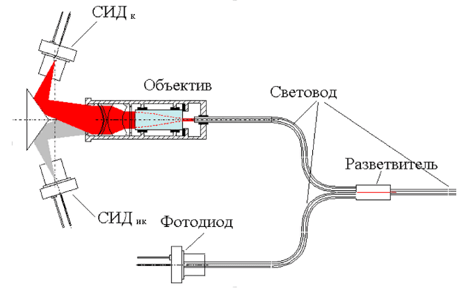
Figure 1 - Optical scheme
The conversion of an optical signal into an electrical occurs through the phenomenon of an internal photoelectric effect in semiconductors. The output current of the photodiode is determined by the formula:
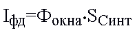
where Iфд is the output current of the photodiode, Фокна is the magnitude of the light flux falling on the photodiode, Scинт is the integrated sensitivity of the photodiode to the signal.
The output signal of the measuring transducer can be considered the voltage value from the output of the pre-amplifier.
Dual-frequency measurement circuit is designed to compensate for disturbances. The output signal in such a circuit is defined as the ratio of the signals of two spectral channels.
Knowing the relationship between partial pressure and saturation, the degree of oxygen saturation of human arterial blood is indirectly determined.
6 Evaluation of metrological parameters
To estimate the instrumental error of the designed measuring instrument, its mathematical model with the structure shown in fig. 2:
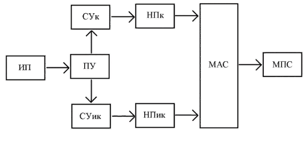
Figure 2 - Block diagram of the device
where ИП is a measuring transducer, ПУ is a preamplifier, СУк is a matching amplifier over the red channel, СУик is a matching amplifier over an infrared channel, НПк, НПик are normalizing transducers of the first and second channels, respectively, МАС is a analog signal multiplexer, МПС is a microprocessor system .
In the process of developing a mathematical model, the processes of setting up electronic devices are modeled using the example of a normalizing converter. As a result of the adjustment, a large part of the systematic error (zero offset) is eliminated, the rest is present as a part of the systematic error that is not excluded. The transmission coefficient is adjusted, the non-excluded part of the multiplicative error is set. The model does not provide absolutely accurate settings for these parameters. The permissible tuning error does not exceed the quantization error of the signal. The instrumental error also includes the errors of the ADC, the computational errors of the scaling algorithms (partial pressure calculations) and the saturation calculation algorithm.
The adjustment error of the partial pressure calculation algorithm was 0.08%, and the saturation calculation was 0.8%.
7 Development of the device design
Using the technology of integrated circuits and optoelectronics, miniature optical sensors sensitive to body analytes in real time have been developed for in vivo analysis of blood gas composition.
Due to significant advances in the development of the communications industry, quite thin optical fibers (optical fibers), high-energy light sources (lasers, light-emitting diodes), and optical radiation detectors appeared.
An optical system for monitoring blood gas composition for resuscitation and surgical departments was developed using a sensor inserted into the patient with a catheter in the radial artery.
The blood oxygen level can be monitored using an intravascular fiber optic catheter. These catheters are already being used [1][2] to monitor the mixed venous blood oxygen saturation during heart surgery and in the intensive care unit.
In fig. 3 shows a fiber optic device for measuring blood oxygen saturation. The device consists of red and infrared LEDs, a photo sensor and a plastic light guide. Plastic optical fibers are adapted to selected wavelengths.
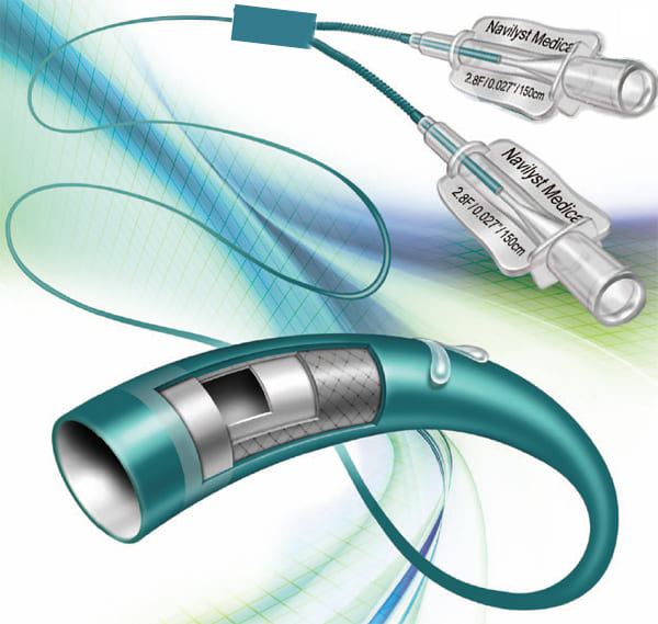
Figure 3 - Optical fiber device for measuring blood oxygen saturation
Conclusions:
In the framework of the research carried out:
- An electronic device has been developed for invasively measuring the content of oxyhemoglobin in human arterial blood.
- In the course of this work, the methods for measuring the degree of saturation of human blood with oxygen were studied, they were analyzed and the most suitable method was chosen.
- A mathematical model for the formation of an analytical signal by the invasive method was chosen.
- The conversion characteristic of the measuring converter of the reflection coefficient (partial pressure) into the photocurrent value of the radiation detector is established.
- A mathematical model of the transformation of partial pressure - saturation is constructed.
- A model of a measuring instrument for estimation of metrological parameters has been built.
- Developed a block diagram of the device. Schematic solutions for structural components are proposed.
- Design elements of the device for invasive measurement of the content of oxyhemoglobin in human arterial blood have been developed.
List of sources
- Основы оптоэлектроники: Пер.с яп.-М.: Мир, 1988.- 288с.; Дональд Дж. Стерлинг. Волоконная оптика: Перю с англ.-М.: Лори, 1998.
- Федотов А.А., Акулов С.А. Измерительные преобразователи биомедицинских сигналов систем клинического мониторинга. –М.: Радио и связь, 2013. – 248 с.
- Принципы мониторинга функции внешнего дыхания. Возможные источники погрешностей при пульсокиметрии[Электронный ресурс]. – Режим доступа: http://kurs.znate.ru/docs/index-149072.html?page=4
- Алейников А.Ф., Гридчин В.А., Цапенко М.П. Датчики (перспективные направления развития): Учеб. пособие / под ред. проф. М.П. Цапенко. – Новосибирск: Изд-во НГТУ, 2001 – 176 с.
- Аналого-цифровые и цифро-аналоговые преобразователи. Справочник. – СПб.: КОРОНА принт.; М.: «Альтекс-А», 2003 – 224с., ил.
- Знакосинтезирующие индикаторы: Справочик/ Под ред. В.П. Балашова – М.: Радио и связь, 1987 – 576 с.: ил.
- Все о резисторах: Справочник. – М.: Горячая линия – Телеком, 1999. ППЗУ – 192 с.: ил. – (Массовая радиобиблиотека; 1239)
- Оптические разветвители в сетях доступа [Электронный ресурс]. – Режим доступа: http://deps.ua/knowegable-base-ru/articles/item/467-opticheskie-razvetviteli-v-setjah-dostupa.html
- Конструкторско-технологическое проектирование электронной аппаратуры: Учебник для вузов / К. И. Билибин, А. И. Власов, Л. В. Журавлева и др. Под общ. ред. В. А. Шахнова– М.: Изд-во МГТУ им. Н. Э. Баумана, 2002 – 528 с.: ил. – (Сер. Информатика в техническом университете.)
- Электронные устройства в медицинских приборах: Учебное пособие / Т. М. Агаханян, В.Г. Никитаев – М.: БИНОМ. Лаборатория знаний, 2005. – 510 с.: ил.
- Полупроводниковые оптоэлектронные приборы: Справочник/ В. И. Иванов, А.И. Аксенов, А.М. Юшин – 2-е изд., перераб. и доп. – М.: Энергоатомиздат, 1988. – 448 с.: ил.