Abstract
Сontents
- Introduction
- 1. The general formulation of the problem
- 2. Review of existing cancer diagnosis on the basis of thermography
- 3. Review of modern methods of processing images termommamografic
- 4. Calculation of the diagnostic features and implementation methods for processing the thermograms
- 5. The structure of a specialized computer system
- Summary
- Sources
Introduction
Computer diagnosis in the modern world is perhaps the most common method of diagnosing the general condition of the body, as well as it's separate organs and systems. The vastness of the current problems of diagnosis and treatment of diseases of the breast is determined by it's scale, prevalence and social significance. Breast cancer — a rather heterogeneous group of malignant neoplasms. Depending on the type of cancer, it's size, position, growth characteristics, presence of metastases and some other parameters will be different and tactics of treatment, and prognosis of the disease.
There are three ways in which we can diagnose breast cancer at an early stage:
- The revision of the breast by the woman.
- Clinical examination.
- Mammography.
In the differential diagnosis of contact thermography plays an important role, namely, to identify breast disease and the formulation of a preliminary diagnosis.
1.The general formulation of the problem
Relevance
To date, breast cancer (BC) women took the leading position, and is the second death, after lung cancer. Therefore, diagnosis of disease at an early stage can significantly reduce mortality in patients after treatment. An important role is played by the means and methods of diagnosis, to determine their capabilities and limits, the search for significant items in mammalogy for mass screening [5]. In the X-ray screening are well known drawbacks: a large number of false-positive diagnoses, the increase in the number of unnecessary biopsies and operations [8‒10]. Some authors have noted a low sensitivity and specificity of the method, the increase in radiation exposure to the breast, which by itself can induce BC [11].
Aims and Objectives
Consider the modern methods of processing digital thermographic imaging in the diagnosis of breast cancer. Building a model according to the thermography of the breast and it's analysis provides an additional criterion for the diagnosis of the disease without adverse effects on the patient's body. Software implementation, based on image processing termomammografic can solve the problem of detection, isolation and analysis, significant for the diagnosis of objects.
The object diagnosis
The object of this study is the mammary gland. In particular, we are interested in breast disease and received during the preliminary diagnosis of this type of thermogram, as shown in fig. 1.a) and below. Thermogram — a temperature map of the area or the whole body to be displayed as an image.
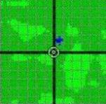
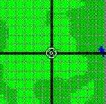
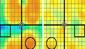
Figure 1 — а) Healthy breast thermogram; b) Thermogram of the right breast cancer.
Expected scientific novelty
The novelty of this project is to construct three-dimensional model of breast cancer on the basis of diagnostic features, and its analysis allows to obtain an additional criterion for diagnosis. This method will enable the differential diagnosis with the help of contact thermography to a new level. Also as part of this work have been identified, such diagnostic indicators such as the local excess of the maximum and the maximum local asymmetry. On this basis, we identify the boundaries of the anomalous zones as shown in figure 1.b).
2. Review of existing cancer diagnosis on the basis of thermography
Thermography is based on the measurement of thermal infrared radiation of the body and gives the true temperature of only the top layer of skin thickness of 2‒3 mm [4]. In medical practice, tested four types of thermal diagnostics: LCD, IR remote, contact thermography and microwave radiometry.
Analysis of the material on the contact thermography has shown its versatility and high efficiency compared with other types of differential diagnosis. Systems based on the thermogram, presented devices such as thermal imaging computer system TV-03K [1], Diagraph DOT-1, thermograph pin digital (TPD_1), which is depicted in figure 2. These systems are similar in principle of the hardware system and intuitive user interface. The most successful in the digital processing is TPD_1 for examination of breast, prostate, bone and joints.

Figure 2 — Appearance of the TPD_1.
The possibility of specialized computer systems (SCS) diagnosis of BC
The advantage of SCS diagnosis of breast cancer by thermography is their suitability for large-scale screening, which involves the ability of these devices in less time and with maximum reliability to obtain temperature maps of the area of the body.
At the same diagnostic methods and devices are absolutely safe for health, both patients and medical staff — a compact, mobile, simple and easy-to-use complexes.
They are able to document the small temperature gradients and distributions, not only on the skin, but also within the body [5], and to fully take advantage of the differential diagnosis (ie, the comparison of thermograms of symmetrical parts of the body) in the visible economy.
3. Review of modern methods of processing images termommamografic
In the region of automation of the thermograms of research should be aimed at achieving the following objectives [4]:
- Expanding the scope and accuracy of diagnostic indicators and analysis of subtle properties of the thermograms.
- Development of integrated assessment methods thermograms.
- Determining the feasibility of using thermography during public examinations and checkups.
- Create a database of thermograms patients.
- Isolation of thermographic signs with subsequent transition to the classification of pathology and diagnosis of computer methods.
- Construction the 3D models for a more in-depth breast diagnosis.
Modern methods of processing the thermograms
Qualitative characteristics of BC diagnostic device, based on thermography are temperature readings of the test section and temperature maps as an image. These characteristics are the primary statistical analysis. There are the following mathematical methods of modeling and processing:
- interpolation and averaging of the temperature;
- edge detection of anomalous zones in the symmetric comparison of temperature on the right and left breast;
- presence of gradients, the confidence intervals of the temperature distribution;
- histogram and isotherm;
- local maximum to the average temperature of the right and left breast;
- maximum asymmetry of the local;
- correlation and scatter plot;
- scanning thermograms and sharpening;
- smoothing filter, contrast, cropping;
- methods Kenny, Sobel, Robert, and other to highlight areas interest [2];
- edge detection of binary object rotation [2];
- build 3D models of breast.
4. Calculation of the diagnostic features and implementation methods for processing the thermograms
The most important role in the diagnosis of breast cancer are diagnostic features. Calculated local maximum to the average temperature of the right and left breast and a local maximum asymmetry are presented in the table 1. The following table 1 shows the area of zones of deviation from the mean temperature of the thermogram for each of the glands, calculated as a percentage of the total area of the thermogram corresponding to the breast.
Table 1. Diagnostic features for localizing areas of hyperthermia
| Parameter | Value, С | S of the scanning spot, cm2 | Quadrant | distance from the nipple, cm | ||||||||||||||||||||||||||
| Max. local to the average temperature of the right breast |
|
|
|
| ||||||||||||||||||||||||||
| Max. local to the average temperature of the left breast |
|
|
|
| ||||||||||||||||||||||||||
| Max. local asymmetry |
|
|
|
|
|
| Zones of deviation from the mean temperature of the thermogram: | Right breast | Left breast |
| The area (more ‒3.0 C) as a percentage of the area of breast thermograms | 4.7 % | 0.0 % |
| (‒3.0…‒2.0 С) | 6.8 % | 0.0 % |
| (‒2.0…‒1.0 С) | 14.0 % | 2.6 % |
| (‒1.0…0.0 С) | 19.5 % | 50.1 % |
| (0.0…1.0 С) | 28.2 % | 46.9 % |
| (1.0…2.0 С) | 26.7 % | 0.4 % |
| (2.0…3.0 С) | 0.0 % | 0.0 % |
| (>3.0 С) | 0.1 % | 0.0 % |
ЗD visualization of the breast
Important for the diagnosis of breast cancer is to build a 3D model in the form of a hemisphere of the breast obtained from a 2D thermogram. Upon receipt of such a model, it becomes possible to hold the points of hyperthermia and hypothermia, perpendicular length equal to the difference of temperatures in the symmetric region of the second breast, as shown in fig. 3 [7].

Figure 3 — Perpendiculars, length in cm C is equivalent to the table
The heat source may be a tumor or inflammation, which have different dimensions and shapes. Many of these perpendiculars visually represents their views. In this fall the perpendiculars from the points that correspond to the temperature difference between the obtained values, ie from the surface of the breast model. The other ends are connected perpendicular to each other and form a figure presented in the figure. In the case of breast cancer should be directed perpendicular to one area (center of hyperthermia or hypothermia), as seen in fig. 4‒5. In the case of mastitis there is no definite direction, and the figure is obtained by the large size. In fig. 4 shows a hypothetical model of the form breast, the first version of the 2D representation, the second — in the form of three-dimensional image. Experimentally, this method will be explored in the thermograms with a known and proven by other means of diagnosis. If a positive result, it is assumed in-depth study of the dependence of the direction perpendicular to the shape and structure of biological tissues of breast.
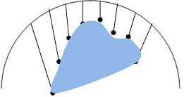
Figure 4 — Model of the breast with the center of hyperthermia
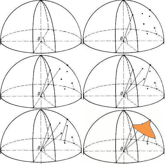
Figure 5 – Model of the breast with the center of hypothermia
(animation: 9 frames, 5 cycles of repetition between frames with a delay 0.5 s., 207 Kb)
5. The structure of a specialized computer system
- The input of data arrives in the form of thermographic cards, with rates of breast temperature of each sensor.
- In the computer diagnostics options are static primary processing, which are formed as a result of diagnostic features.
- Diagnostic characters are entered into the protocol and are used for visualization of the breast and the diagnosis.
- The result — an effective and accurate differential diagnosis and the possibility of using the system for mass screening.
Expected results of a computer system designed
- increase in sensitivity and specificity of contact thermography;
- reducing time-consuming;
- provision of system options to preserve the diagnostic results.
Summary
The study examined methods of calculating the diagnostic signs of breast cancer — deviations from the normal temperature and the local asymmetry. Due to the need for the diagnosis of breast cancer to build three-dimensional model of the breast. Software implementation and testing in the hospital showed that the methods for isolating areas of pathology, based on thermography are efficient, reasonably accurate and can be successfully used in the differential diagnosis of breast disease.
Sources
- Tkachenko А.Y. Clinical Thermography (overview of the main features) / А.Y. Tkachenko, M.V. Golovanov, А.М. Ovechkin — Nizhny Novgorod, 1998 y.
- Titova A.Y. Digital image processing in mammography. — Proceedings II of the All-Ukrainian scientific-technical conference of students and young scientists (Volume 3) — Donetsk, 2011 y.
- Приходченко В.В. Применение контактного цифрового термографа ТКЦ-1 в диагностике заболеваний молочных желез — Руководство для врачей. / В.В. Приходченко, Ю.В. Думанский, О.В. Приходченко, В.А. Белошенко, В.Д. Дорошев, А.С. Карначёв — Донецк, 2007 г.
- Розенфельд Л.Г. Дистанционная инфракрасная термография в онкологии. Онкология. / Л.Г. Розенфельд, Н.Н. Колотилов — 2001 г. — т. 3., № 2‒3., с. 103‒106.
- Белошенко В.А. Комплекс аппаратуры для ранней диагностики онкологических заболеваний методом контактной цифровой термографии. / В.А. Белошенко, В.В. Приходченко, В.Д. Дорошев, А.С. Карначёв — Наука та інновації № 5. — 2007 г.
- Cockburn W. Nondestructive testing of humanbreast. // www.breastthermograpy.org/SRIE.htm.
- Титова А.Ю. Специализированная компьютерная система обработки термоммамографических изображений. — Сборник материалов II Всеукраинской научно-технической конференции студентов, аспирантов и молодых ученых (Том 1). — Донецк, 2012 г.
- Пономарев И.О. Медицинский скрининг — проблемы, перспективы и возможности применения в онкологии // Онкология. — 2001. —Т. 3, № 2‒3. — c. 203‒206.
- Орлов О.А. Медицинские и экономические аспекты маммологических профилактических осмотров женщин // Казанский мед, журнал. — 2001. — Т. 82, № 5. — c. 388‒391.
- Семиглазов В.Ф. Промежуточные результаты программы Россия (Санкт-Петербург) / ВОЗ по оценке эффективности самообследования молочных желез/ Вопросы онкологии. / В.Ф. Семиглазов, В.М. Моисеенко, С.А. Проценко и др. — 1996. — Т. 42, № 4.— c.49‒55.
- Skrabanek P. Shadows over screening mammography // Clin.Radiol. — 1989. — 40, № 1. — Р. 4‒5.
*This master's work is not completed yet. Final completion: December 2012. The full text of the work and materials on the topic can be obtained from the author or his head after this date.
To the beginning
