
Abstract
Content
- 1. Theme urgency
- 2. Goal and tasks of the research
- 3. Preliminary preparation
- 4. Photogrammetric System
- 4.1 Camera for shooting
- 4.2 Description of installation
- 5. Image processing
- 5.1 Rationale for the ERDAS software to build digital models
- Conclusion
- References
1. Theme urgency
In many fields of science and practice there is the problem of determining the geometric characteristics of the surface of the biological objects. One such area is the medicine. In particular, characterization of the human body surface and changing their time necessary for diagnosing and monitoring the disease [1].
Designed to date methods of determining the geometric characteristics of the surface of the biological objects do not meet modern requirements, because either give very little information, but very simple methods, or require time‐consuming to carry out measurements using expensive equipment and processes resulting from these measurement data.
Obvious urgency of developing such a method of determining the geometric characteristics of the surface of the biological objects, which must have a high efficiency, sufficient precision and relatively low cost. Moreover, this method must be safe for the test object.
Such requirements are fully meet the photogrammetric techniques, since these methods:
– Are non‐contact and, unlike X‐ray method is absolutely safe for the human body;
– The safety of these methods allows for repeated shooting with any interval of time;
– Allow us to construct a three‐dimensional mathematical model of the surface of the entire body, not just its fragments;
– Can take pictures of the body surface with very high accuracy (up to tenths of a millimeter);
– It can be implemented with low‐cost consumer digital cameras and standard software systems for digital photogrammetric processing of images.
Due to its high accuracy, versatility and performance of tasks information technologies could not find application in medicine, particularly in dentistry. Even the term "Dental Informatics" and "computer dentistry."
Computer processing of graphic information quickly and carefully examine the patient and to show the results both to the patient and other professionals. [2]
Difficulties in measuring the parameters of parts of the adjacent tooth jaw appear in determining the relative position of the inner surface of the adjacent parts. This issue is very important for dentistry, where accurate measurements with respect to the upper and lower teeth, and the relevant provisions (occlusion) are necessary for effective treatment. Therefore, more and more is offered Photogrammetric equipment positioning for the analysis of the jaw. Computer processing of graphic information quickly and carefully examine the patient and to show results both to the patient and other professionals. A dentist can examine the relative position of the teeth and measure the distance between the teeth in different locations of the jaw. Also, this method allows you to virtually explore how the prosthesis will interact with the other teeth.
2. Goal and tasks of the research
Goal: To study the use of specialized cameras to build a digital model of a dental jaw.
Tasks of the research:
1. Creating a photogrammetric system for shooting
2. Surveying installation
3. Study of the software used in the work for the construction of a digital model
4. Building a digital model of a dental impression of the jaw
The object of the research is to build a digital model of a dental impression of the jaw, and the subject – non‐specialized digital camera.
Scientific novelty consists in the use of specialized cameras for shooting pictures on which to build a digital model.
3. Preliminary preparation
For successful treatment of teeth, it is important to have information about their relative position and know the distance between the teeth. The definition of these distances – a task for the dentist, as today is not developed an electronic system to identify them. Dentist receives information about the position of the teeth, the presence (or absence) of contact between the teeth. This information is obtained by means of thin sheets of color paper, noting the location of contact on the teeth. According to the information already produced a dental impression of the jaw, which is used for shooting and based on which build a digital model of the jaw.
Shooting impression is sequentially moving the camera and fixing the image in each transfer (Fig. 1). Slepok must be located in a stationary fixed position, that will considerably simplify processing of of shots and the construction of of digital model of. To cast remained motionless during the shooting must be put on a flat surface and secure.
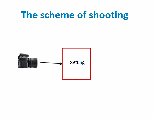
Figure 1 – The scheme of shooting
(animation: 5 frames, 10 cycles of repetition, 31.7 KB)
To construct a three‐dimensional model using the software, and not enough to have a cast chamber, a problem arises in the absence of the coordinate system for processing images need a set of points with known three‐dimensional coordinates [3]. Points should have different coordinate Z. At this stage, the problem is to fix those reference points and their location. In addition, the point to be measured in a coordinate system, which is also a problem, since it is necessary to digitize this coordinate system.
In the process of taking a picture of the jaw model and all reference points must be sufficiently lit, which also need to be addressed in the preparatory phase.
4. Photogrammetric System
The procedure of constructing a digital model includes several stages. In the first stage we made a plaster model of the jaw. Then, the three‐dimensional model of a jaw generated by using a system based on photogrammetric cameras and installation. The camera model of the jaw, forming a set of partial models, which are then merged using special software. The method uses a set of control points. Images jaw formed photogrammetric system and 3D coordinates of the reference points are designed to build a digital model.
4.1 Camera for shooting
The work will be studied camera Nikon D5100 [4]. This is a digital single‐lens reflex camera. Taking the camera performs at the highest magnification, that is, when the maximum focal length, in which, as we know, the manifestation of optical distortion is minimal.
Key features Nikon D5100
• CMOS‐sensor with a resolution of 16.2 megapixels;
• Wide range of ISO – 100 to 6400 + expandable to ISO 25600. A high level allows the use of faster shutter speeds in poor lighting conditions, thus avoiding blurring;
• Swivel LCD screen with a diagonal of 3 inches and a resolution of 921,000 pixels;
• The magnification factor is the lens focal length of 1.5.
Formats:
• DCF 2.0 (Design Rule for Camera File System)
• DPOF (Digital Print Order Format)
• Exif 2.3 (Exchangeable Image File Format for Digital Still Cameras)
• PictBridge
4.2 Description of installation
The paper envisages the creation of installation, which is the backbone of the jaw and the dental cast (Fig. 2).
In this paper, we propose the following basic network:
1. Glass that is used to calibrate stereocomparator with mesh size – 5 mm and precision coating lines less than 0.05 mm, which makes it possible to control the position of the object and reference points, as well as to carry out a rough orientation in the pictures.
2. Pegs with attached on them trademarks (anchor points). The accuracy of the creation better than 0.1 mm.

Figure 2 – Removable installation
The figure shows that the mold is on the glass coated with a grid. In addition impression taking are also present of the brand, which are mounted on carved on the size pegs. Pegs have a different the excess of. 3 counterbalanced point were taken on the very glass.
5. Image processing
Was last is executed trial shooting installation. Schematic capture is shown in Figure 1. The camera consistently moves relative to your subject and capture images, while respecting the images overlap (60%) [5]. The total number of pictures – 5.
The results of the measurements on the pictures was built focused and scaled geometric model, ie a set of stereo model, oriented in a single coordinate system.
The measurements were performed in photogrammetric complex Delta (fig. 3).

Figure 3 – Measurements
In as binders points of were used the point of at the intersection of grid of coordinates, additional of the brand and marks on the the very the impression.
Equalizing of measurements is performed in BlockMSG. If the adjustment is necessary to obtain accuracy better than 0.5 mm. When the adjustment error was found to be 3 mm, which does not satisfy the required accuracy.
Therefore, has been executed the second shooting with exception of previous errors. To improve the accuracy, it was decided the use of the software product ERDAS to build a digital model.
5.1 Rationale for the ERDAS software to build digital models
ERDAS Imagine – The full‐featured system for work with snapshots [6].
Rapidly growing information requests modern society dictate the the need for adequate means of obtaining and processing the necessary data. It has caused development of many directions of software industry, in particular information processing systems. The first violin in this party plays a the company ERDAS Inc. which is since 1978 provides a thousands of users a powerful software for raster of geoinformation systems and image processing. Right‐flank in the line of software products company's – a package of ERDAS Imagine – full‐featured system created for solving applied problems today and tomorrow.
Understandable a graphical interface and powerful means of processing a spatially distributed information provide a fully functional environment for solutions a wide spectrum of applied tasks. Graphically related the window viewing (windows Viewer'a) allow simultaneously display and carry out the analysis and various transformation of information. Special format provides quick a conclusion of large of graphic files with high resolution. Context‐sensitive tooltip organized in a hypertext help‐system as well as extensive and well‐written printed manuals properly provide a "the rear of".
With the help of ERDAS Imagine you can quickly to receive access to the most of diverse information that the gives unlimited possibilities for the analysis of and management of databases. Of available funds you can get information as the on a territorial object so‐and significance of of each pixel the image and associated with him an additional information to use various means and opportunities visualization, in particular, scaling.
ERDAS Imagine allows you to create digital models. In this case all operations are performed programmatically without the use of the special photogrammetric apparatus. The module Digital Ortho in ERDAS Imagine uses a phototriangulation for the creation of digital model from snapshots. The resulting a digital model of can use to create of three‐dimensional perspective image [7].
The software of American firm ERDAS Inc. It provides a all complex of funds needed for effective use of of data filming in any possible areas of their application. It includes basic kit and expansion modules. Allows you to improve the quality and improve the accuracy of the image transform the snapshots tethering snapshots to each other classify objects to filter out "noises" to synthesize the multispectral images to simultaneously analyze raster and vector the information create high‐quality professionally decorated maps and much more. A variety of means of image analysis let you receive the rich information for a plurality of applied tasks.
Software products of firm ERDAS combine in itself functions of raster of geoinformation system (GIS) and systems for image processing and represent a modular products are running on many computer platforms.
Conclusion
As a result, the work will be justified use of specialized cameras to build a digital model of a dental jaw. These digital models constructed with the use of the camera can be further used a dentist to correct the defects, selection of the optimal variant of treatment of a patient.
In writing this essay master's work is not finished yet. The final completion – December 2015. The full text of work and materials on the topic can be obtained from the author or his manager after that date.
References
- Абдульмунеам А. М. «Разработка и исследование фотограмметрических методов определения геометрических характеристик поверхности биологических объектов» [Электронный ресурс]. – Режим доступа: http://www.dissercat.com/content/razrabotka-i-issledovanie-fotogrammetricheskikh-metodov-opredeleniya-geometricheskikh-kharak
- Семинар "ДОПОЛНИТЕЛЬНЫЕ МЕТОДЫ ОБСЛЕДОВАНИЯ В СТОМАТОЛОГИИ" [Электронный ресурс]. – Режим доступа: http://xn----btbhd8ap0b6a.xn--80ao21a/questionary/view/82.html
- Буров М. И. Практикум по фотограмметрии / Буров М. И. – М.: НЕДРА, 1987. – 302 с.
- Полный обзор фотоаппарата Nikon D5100; Технические характеристики и цены на Nikon D5100 kit и body [Электронный ресурс]. – Режим доступа: http://digitalservis.ru/nikon/678/nikon-d5100.html/
- Бирюков В. С. «Цифровые снимки в фотограмметрии» / Геодезия и картография. – 2000. – N10. – С. 33‐36 [Электронный ресурс]. – Режим доступа: http://www.stereo-pixel.ru/docs/pressa/1/dig_imgs.htm
- Дистанционное зондирование [Электронный ресурс]. – Режим доступа: http://loi.sscc.ru/gis/gt/9.htm
- Создание ЦСММ с использованием стереопар в ERDAS IMAGINE [Электронный ресурс]. – Режим доступа: http://gis-lab.info/qa/stereo-erdas.html .