
Abstract on the topic of the final work
Content
- Introduction
- 1. Relevance of the topic
- 2. Purpose and objectives of the study, planned results
- 3. The structure of the developed biotechnical system
- 3.1 Block diagram and its components
- 3.2 System characteristic
- 3.3 Provision levels
- 4. Characteristics of the object of study
- Conclusions
- List of sources
Introduction
This article presents a mobile system for analyzing the patient's muscle tone. The system consists of a mobile device, such as a smartphone or tablet, equipped with sensors to measure muscle activity. Sensors are placed on the patient's skin at strategic locations and are used to collect data on the patient's muscle activity. This data is then analyzed using machine learning algorithms to determine the patient's muscle tone. The system is designed to be portable and easy to use, making it a useful tool for physical therapists and other healthcare professionals who need to assess muscle tone in their patients. The system can also be used to study and track changes in muscle tone over time.[1]
1. Relevance of the topic
The assessment of muscle tone is an important aspect of physiotherapy and rehabilitation, as well as in the field of sports medicine. It is used to assess the level of muscle activity and identify any abnormalities or imbalances in muscle tone. It is also used to monitor the progress of a patient's recovery and track changes in muscle tone over time.
A mobile system for analyzing muscle tone is particularly relevant as it allows healthcare professionals to perform assessments in a variety of settings, including in the clinic, at home, or even remotely. This can be especially helpful for patients who have mobility issues or live in remote areas, as it eliminates the need to travel to the clinic for an assessment.
In addition, using machine learning algorithms to analyze muscle tone data can improve the accuracy and consistency of estimates, as well as detect subtle changes in muscle tone that can be missed by traditional methods.
In general, a mobile system for analyzing muscle tone can provide a more efficient,a convenient and accurate way for healthcare professionals to assess and monitor their patients' muscle tone and can help improve patient outcomes.
2. Purpose and objectives of the study, planned results
Purpose - to develop a hardware-software complex for the analysis of muscle tone to improve the efficiency of decision-making in the treatment of patients with neuromuscular diseases
Main research objectives:
— Development of device hardware: design and creation of electronic circuits, selection and integration of electronic components, production of printed circuit boards and mechanical parts;
—Software development: creating a program that will process and display the captured signals, as well as provide communication with other devices (for example, with doctor's monitors);
—Implementation of signal analysis processing algorithms and software.
Research object: When developing a biotechnical system for analyzing a patient's muscle tone, the object of research will be the biotechnical system itself, including an EMG recording device, an analyzer and software for data processing and analysis.
Subject of study:
— Information support;
— Methodological support;
— Tooling;
—Software and algorithmic support;
— Metrological support.
Planned scientific results: Obtaining new knowledge about the functioning of the human nervous system and muscles, as well as methods for diagnosing and monitoring them. A new understanding of the technologies for collecting and processing EMG signals can also be obtained.
Planned practical results: The possibility of using the device in medical institutions for the diagnosis and monitoring of various diseases of the nervous system and muscles. Also, the device can be used in operations on the nervous system and muscles to continuously monitor the patient's condition.
3. The structure of the developed biotechnical systems
This BTS is diagnostic, based on physiological methods based on the registration and analysis of bioelectrical potentials of muscles and peripheral nerves. At the same time, depending on the objectives of the study, both voluntary and stimulation-induced activity of the neuromuscular apparatus is evaluated.
3.1 Block diagram and its components
The structure of the developed biotechnical system is shown in Figure 1: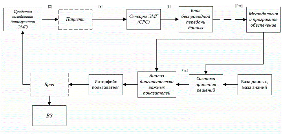
Figure 1 - Structural diagram of the developed BTS
Figure 1 shows the structure of contactless monitoring, where:
– Signal recording tools (EMG, which picks up a signal using electrodes, based on the difference in electrical potential between two points);
– Means for processing medical signals and data (in this case, it is a methodology and a program);
– Medical decision support system (coordination with the medical information system is underway)
- Analysis of diagnostically significant indicators (such as H-reflex, F-wave, M-response, myotatic reflex, used in the assessment of nerve damage);
VZ - medical opinion.
[X] – patient's reaction to the test impact;
[Y] – set of biomedical signals reflecting the patient's response to the test effect [X];
[S] is a set of signals recorded by sensors and electrodes ([S] = [K][Y], where [K] is a linear operator that reflects the sensitivity of the sensors, and is also the gain of the amplifiers);
[Pts] – Indicator of the current state of the patient.
3.2 System characteristics
System control loops include setting measurement parameters, managing sensors, and displaying measurement results on the device. The system can also have the function of automatically alerting medical personnel about deviations from the norm in the patient's health parameters.
The communication channels of the system may include a wireless connection to a hospital LAN or the Internet to allow medical staff to monitor a patient's condition remotely. The function of recording and storing data can also be implemented to enable the information to be analyzed in the future.
System communication channels provide data transfer between various devices and system components. They can be implemented using various technologies, depending on the required speed, distance and transmission security.data.[2]
Some examples of information exchange channels that can be implemented in the system:
- Wireless connection: Using technologies such as Bluetooth or Wi-Fi, you can realize a wireless connection between sensors and a portable device. A wireless connection to the hospital's LAN or the Internet can also be implemented to enable remote patient monitoring.
- Wired connection: A wired connection can be made between the sensors and the handheld device using cables and adapters. This option may be suitable if you want to ensure high speed and accuracy of data transfer, as well as avoid possible interference in the system operation associated with the use of wireless technologies.
- Integration with other systems: The system can be integrated with other medical systems such as intensive care monitoring systems, infusion management systems, etc. For this, various interfaces and data exchange protocols can be used, such as HL7, DICOM, etc. Integration with other systems enables data sharing and improves treatment efficiency, as well as reducing the risk of data entry errors.[3]
Depending on the required functions and characteristics of the system, information exchange channels can be implemented as a combination of different technologies and interfaces. In the master's thesis, it is planned to develop: information, methodological, instrumental, software-algorithmic and metrological levels of support (components).[4]
3.3 Security levels
To develop this system, the following levels of support should be developed:
1. Information support:
- Database for storing information about patients, EMG signal measurements, treatment, etc.;
- Interfaces for entering, editing and displaying information;
- Database management system to ensure data security, integrity and consistency.
2. Methodological support:
- Guidelines and instructions for using the system for medical professionals and patients;
- EMG signal processing and analysis techniques;
- Knowledge base about pathologies and their treatment
3. Tooling:
- Equipment for taking the EMG signal (for example, an electrocardiograph or an electromyograph);
- Software for EMG signal processing and analysis;
- Tools for visualizing and displaying results (for example, graphs and charts).
4. Metrological assurance:
- Standards and specifications necessary to ensure measurement accuracy;
- Means of calibration and verification of equipment;
- Metrological documentation management system.
5. Software and algorithmic support:
- Operating system and required software;
- The program code that ensures the operation of the system;
- Software version control system and ensuring compatibility with various operations
The structure of the developed system provisioning level may include the following components:
1. Data collection and processing module:
- Equipment for taking EMG signal;
- Software for EMG signal processing and analysis;
- Interfaces for data input and display.
2. Information storage and processing module:
- Database for storing information about patients, EMG signal measurements, treatment, etc.;
- Database management system
3. Visualization and analysis module:
- Graphing and charting functions;
- Data analysis functions that allow you to identify trends and patterns;
- Interfaces for displaying analysis results.
4. Quality Management Module:
- A quality management system that allows you to monitor the compliance of the system with requirements and standards;
- Tools for auditing and analyzing the quality of the system.
5. Security module:
- Functions of data encryption and protection against unauthorized access;
- System security monitoring functions;
- Data backup functions.
6. Project management module:
- Software version control system;
- Task management system
- Tools for planning, controlling and tracking development progress;
- Documentation management system and regular project status reports.
These components can be developed as independent modules that can be linked and integrated into the overall system. When developing the level of support, one should take into account the needs and characteristics of medical activities, as well as the requirementsto the safety, quality and compatibility of the system.
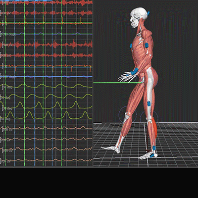
The object of study in this system is the work of the human nervous system and muscles, as well as the possibility of their diagnosis and monitoring using an EMG device. This object is characterized by the following characteristics[5]: 1. Dynamic nature: the work of the human nervous system and muscles changes over time, so continuous monitoring of the state is required. 2. Complexity: The human nervous system and muscles are made up of many different elements that interact with each other. This makes it difficult to understand their work and find ways to diagnose and monitor. 3. Need for Accuracy: Mistakes in diagnosis and monitoring can lead to serious health consequences for the patient.[6] As a device is developed, the mathematical model is an important tool for analyzing and understanding how the nervous system and muscles work. It allows you to simulate the operation of these systems and explore their behavior under various conditions. For example, in this work, one of the functions of the device is continuous monitoring of the state of the nervous system and muscles during operations, for the analysis and processing of data collected by the device, a mathematical model can be used based on the use of functions that describe the dependence of electrical signals on time . This model can help in determining the required signal level for the normal functioning of the nervous system and muscles, as well as in determining the abnormal level of this level, which may indicate a violation of the system and the need for intervention. A typical EMG recording is shown in Figure 2.
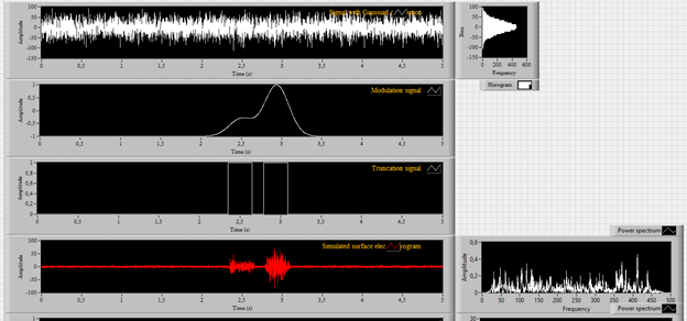
To estimate the spatio-temporal parameters of electrical activity, methods such as frequency, spectral-coherent and correlation analysis are widely used. Frequency analysis allows you to decompose a complex curve of electrical activity of the brain into component frequency components, and obtain a spectrum of bioelectrical brain oscillations, highlight and quantify the frequency components of EMG.[7] Spectral analysis determines not only the severity of various rhythmic components in a complex EMG, but also their ratio, allows you to obtain information about the total power of the EMG spectrum and its frequency components. Typically, the results of the analysis are presented in the form of graphs of power spectral density, graphs of amplitude spectra, topographic maps, as well as various quantitative indicators in tabular form. Coherent analysis determines the statistical relationship between the fluctuations of biopotentials occurring in different parts of the brain, which reflect the degree of synchronism of EMG changes at two different points in the frequency domain (for nearby and distant brain regions within each of the hemispheres and for the same-name brain regions of the right and left hemispheres), and cross-correlation - in time. Let's simulate the EMG signal using a mathematical model based on the use of functions that describe the dependence of electrical signals on time, for this we use the LabView software package. The result is shown in Figure 3. Figure 3 - Results of experimental studies 1. Information support: 2. Methodological support: 3. Tooling: 4. Metrological assurance: 5. Software and algorithmic support: 6. Development and improvement of EMG signal processing and analysis algorithms: 7. Development of integration functions with other systems: 8. Development of control functions 9. Developing Reminders and Automation Functions: 10. Develop interactive learning features: 11. Development of social integration features: 12. Development of personalization features: 13. Development of analytics and reporting functions: 14. Development of security features:4. Characteristics of the object of study
Conclusions
To improve the developed mobile system for EMG signal acquisition and intraoperative monitoring, the following areas of improvement can be considered:
• Development of an intuitive user interface;
• Ensuring the security of information stored in the system;
• Development of mechanisms for processing and analyzing data obtained using the system.
• Development of methods and guidelines for working with the system;
• Training of users to work with the system;
• Conducting seminars and trainings for specialists using the system.
• Development and improvement of hardware for removing the EMG signal;
• Ensuring system compatibility with various models of medical equipment;
• Development and improvement of algorithms for working with hardware.
• Ensuring the accuracy of measurements taken with the system;
• Development and improvement of system calibration algorithms;
• Carrying out regularmeasurement accuracy checks.
• Development and improvement of system software;
• Development and improvement of data processing and analysis algorithms;
• Ensuring system compatibility with various operating systems.
Additionally, you can consider the following areas for improving the developed system:
• Improving the accuracy of determining cardiac arrhythmias;
• Development of algorithms for determining the severity of rhythm disturbances;
• Development of algorithms to determine the possible causes of rhythm disturbances.
• Integration with electronic medical record systems;
• Integration with monitoring systems for other parameters (eg blood pressure, respiration);
• Integration with systems for planning and managing medical procedures.
• Development of functions for managing the parameters of the system operation (for example, the frequency of polling the EMG signal, the frequency of updating information);
• Development of hardware management functions (for example, turning the device on/off, controlling the parameters of the device);
• Development of functions for managing access to the system (for example, managing access levels, managing access rights).
• Development of reminder functions for the need for medical procedures;
• Development of reminder functions for the need to take medication;
• Development of functions for automating routine processes (for example, automatic sending of a report
• Development of functions for automating routine processes (for example, automatic sending of reports to the mail or SMS notifications about the need to take medications).
• Development of online learning functions for system users;
• Development of interactive modules to better understand the operation of the system and the principles of EMG signal processing;
• Development of user knowledge testing functions.
• Development of functions for sharing the system with other medical institutions;
• Development of data exchange functions with other medical systems;
• Development of functions for exchanging information with medical professionals and patients through social networks.
• Development of functions that allow you to customize the system for specific users;
• Development of functions that allow you to save the history of measurements and analyze changes over time;
• Development of features that allow individualized recommendations and treatment plans for each user.
• Development of reporting functions for different levels of management;
• Development of data analysis functions to identify trends and patterns;
• Development of functions for constructing graphs and charts that allow you to visually explore the data.
• Development of functions for data encryption and protection against unauthorized access;
• Development of system security monitoring functions;
• Development of data backup functions.
List of sources