
Abstract
Contents
- Introduction
- 1. Relevance
- 2. Purpose and objectives of the study, planned results
- 3. Types of electrode for electroencephalogram
- 4. Simplified structural diagram of the device
- 5. Types of artifacts on the EEG
- Conclusion
- List of sources
Introduction
Today electroencephalography (EEG) is an available non-invasive method of estimating the functional state of the human brain. This method has established itself as an effective diagnostic tool and is currently widely used in clinical practice.
Nevertheless, modern encephalographs cannot be called cheap, and domestic developments have not been updated for a long time. The task of this work is to design a domestic modern, inexpensive and portable single-channel electroencephalograph, taking into account the subtleties of electroencephalography and interference arising during the study.
1. Relevance
Electroencephalography (EEG) of the brain is a diagnostic method by which the brain is examined It is the most accessible method of determining the condition of the brain, its activity and changes in the condition of cells under various stresses. Thanks to the use of modern technology, the electroencephalograph provides accurate information. This diagnostic method provides an opportunity to reflect the pattern of active activity of all brain systems. The EEG accurately and clearly shows one of the important parameters of the nervous system's activity. This factor is called "rhythmicity property". It reflects the coordinated work of different structures and departments in the brain.
Electroencephalography (EEG) can detect the following abnormalities:
- Consequences of craniocerebral injuries and surgical operations;
- Epileptic disorders;
- Новообразования головного мозга, доброкачественные и злокачественные;
- Brain tumors, benign and malignant;
- Encephalopathies and other diseases of the brain and CNS;
Electroencephalography (EEG) is important in the diagnosis and treatment of various neurotic disorders (depression, insomnia, hysteria, panic attacks, etc.).
2. Purpose and objectives of the study, planned results
The purpose of the work is to substantiate and set technical requirements for the development of an electronic device for monitoring the biological rhythms of the human brain.
As part of the master's work is planned to obtain relevant scientific results in the following areas:
- Review and analysis of existing electroencephalographs.
- Development of a design specification for an electronic EEG signal processing unit.
- Rationale and development of the block diagram of electronic EEG signal processing.
3. Types of electrode for electroencephalogram
A bioelectric electrode is a device used for taking bioelectric potentials, which has a current-carrying surface and output elements. The current-carrying surface is the part of the electrode surface that comes into contact with a bioelectric object directly or through a contact substance and provides for taking bioelectric potentials or connecting the object to a neutral terminal. In addition, a bioelectric electrode has a supporting surface, which is the part of the electrode surface that performs a supporting function when attaching the electrode to the bioobject. Electrodes are used to convert ionic potentials into electronic potentials (voltages and currents in conductors of the first kind). Bioelectric electrodes are classified according to various characteristics, the main of which are the peculiarities of participation of electrodes in taking biopotentials, the frequency of use, the duration of contact with the bioobject in one study, the nature of the source of the bioelectric field under study, the place of application or insertion, the method of keeping in contact with the surface of the bioobject, and some others. Depending on participation in biopotentials withdrawal, the following types of electrodes are distinguished. Potential electrode - a withdrawal electrode contacting a part of a bioobject located in the electric field of the organ under study; zero electrode - a withdrawal electrode contacting a part of a bioobject in which the electric field potential of the organ, tissue, cell under study tends to zero; neutral electrode - an electrode not involved in taking bioelectrical potentials, connected to the neutral terminal of the measuring instrument.
Depending on the source of the bioelectric field under study, the electrodes are divided into the following basic types:
- electrocardiographic and electroencephalographic, applied without disturbing the skin and bony coverings;
- electrocorticographic - for taking biopotentials from the surface of the exposed cerebral cortex;
- electromyelography - to study the biopotentials of the spinal cord;
- electromyography - for taking biopotentials of muscle fibers;
- electro-oculographic - to capture the biopotentials produced by the eyeball movement;
- electroretinographic, applied to the retina;
- electrogastrography - to study biopotentials caused by the electrical activity of the stomach;
- electrocochleography - for recording of biopotentials caused by the activity of structures of the outer, middle and inner ear.
According to the place of application or insertion, there are skin electrodes, corneal electrodes, electrodes for dissected organs, cavity electrodes for insertion into natural body cavities, intratissue electrodes and microelectrodes whose current-carrying surface is designed to remove the electric potentials of the cell and its structures. The first three types of electrodes refer to surface electrodes. Intracavitary electrodes, in turn, are subdivided according to the place of insertion into the body.
Types, sizes and some design features of electrodes used in various types of electrophysiological studies are defined by standards. The material and structure of electrodes are not specified by the standard. Standards also stipulate a number of important electrical parameters of electrodes: electric potential difference between two electrodes in the lead, potential difference drift, noise voltage, total electrical resistance, methods and means of testing electrodes.
There are several types of EEG electrodes used in electroencephalography, which differ both in shape and in the way they are attached to the head:
- Contact overhead non-adhesive electrodes, which are attached to the head by helmet-mesh webs;
- Bridge electrodes;
- Adhesive electrodes;
- Basal electrodes;
- Needle electrodes;
- Cup electrodes;
- Multi-electrode needles.
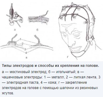
Figure 1 – Types of electrodes for EEG
Modern EEG machines are equipped with electrode helmets with built-in cup electrodes. Along with helmets, it is recommended to use mesh helmets for fixing two types of electrodes: cup or bridge electrodes.
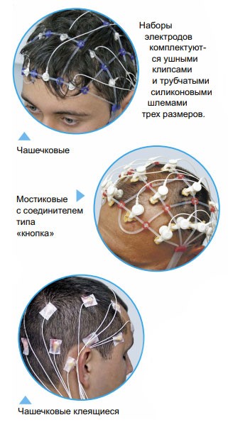
Figure 2 – Cup and bridge electrodes
4. Simplified structural diagram of the device
Electrical potentials that we will be capturing have a small amplitude, normally it is 50-150 µV. Therefore, we must amplify the signal as qualitatively as possible, for this purpose we will use amplifiers with a gain of 20-100 thousand. It is necessary to take into account that during registration powerful electromagnetic fields created by any electrical devices around will act on the signal being taken. The signal taken from the head surface will be influenced by interference in the form of in-phase voltage. To avoid this voltage, differential amplifiers are used, which eliminate the voltage acting equally on both inputs and amplify the difference voltage.
A modern electroencephalograph is a multi-channel recording device that captures the electrical activity of the brain by means of a large number of electrodes placed on the subject's head. It is important to position the electrodes correctly because the potentials taken from different points are different. There are two types of electroencephalogram recording: monopolar and bipolar. During a monopolar recording, the signal is electrically active only relative to some neutral point (earlobe). The bipolar method involves a potential difference between electrodes that are at electrically active points. We will shoot the signal with the monopolar method, because it allows us to analyze the contribution of a specific area of the brain.
There are certain requirements for the material of the electrodes, they must not polarize during signal acquisition. Due to the contact of the skin electrode, ions accumulate on the electrode due to electrochemical processes. This process leads to distortions as a constant component is added to the signal being captured. The best material in terms of reliability is silver. If polarization occurs, the silver electrode is chlorinated, which causes a layer of silver chloride to appear on the surface of the electrode. An electrode paste or solution is also used for better contact. Dry electrodes, such as pin electrodes, for contact on the scalp are becoming more common nowadays.
From the head, the electric potential is fed through the electrode to the input of the amplifiers, which provide a reduction of the input resistance, which is formed by the skin-electrode contact. The number of amplifiers corresponds to the number of electrodes. The electrodes are connected to the device due to the input box, which contains numbered sockets, and can select a particular pair of electrodes, between which will be removed. Also in the box there is a neutral electrode connected to the instrument ground, by which we equalize the potentials of the human body and the amplifiers. The lower the resistance under the neutral electrode, the better the potentials are equalized, consequently, there is less in-phase interference, affecting the studied signal.
Structural diagram of the portable encephalograph is shown in Figure 3. The electroencephalograph circuit consists of two parts - analog and digital. The analog part is a signal received from the input electrodes, each of which is connected to a preamplifier, to provide lower input skin-to-electrode resistance. The signal then goes to the amplifier. The amplified signal goes to the ADC and then to the microcontroller, which filters the signal from noise - this is the digital part of the circuit.
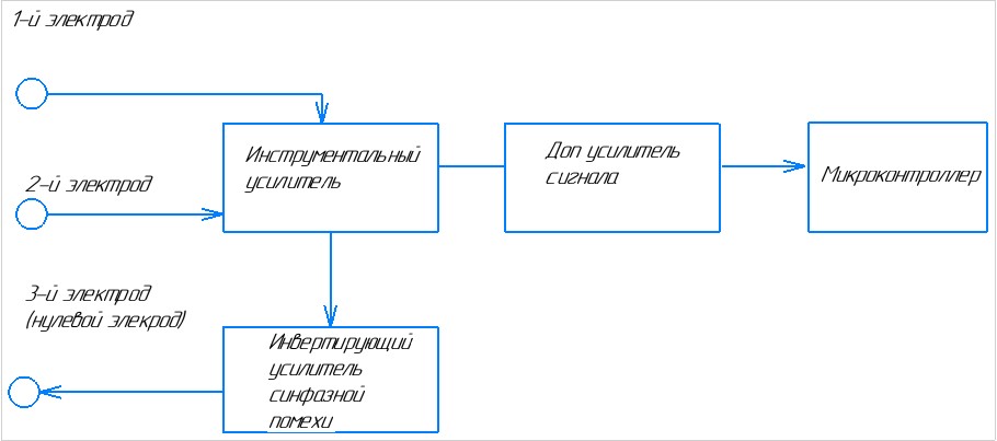
Figure 3 – Generalized device schematic diagram
5. Types of artifacts on the EEG
Modern electroencephalographs have high sensitivity and good interference immunity, which allows recording high-quality EEG practically under any conditions. However, registration of stray signals or noise is still possible, since any EEG complex eventually registers electrical activity, and electrical activity of the brain is much lower in amplitude than, for example, heart and muscle activity.
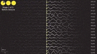
Figure 4 – Example of EEG recording
There are several types of typical artifacts in EEG recording:
Physiological artifacts
- ECG artifact. Since the electrical activity from the contraction of the heart muscle is much higher in amplitude than the activity of the brain, it is often possible to see pronounced sharp peaks from QRS complexes on the EEG. The ECG artifact is particularly evident if one of the electrodes is placed in close proximity to a vessel. The main danger in the ECG artifact is that it can be mistaken for spike activity. Therefore, it is recommended to record one additional channel of ECG when performing EEG examinations.
- EEG artifact. This type of artifact is probably the most common in EEG recording, especially in the open-eye test. This artifact is caused by eye movements of the subject and is most pronounced in the frontal leads. Sometimes this artifact can be mistaken for slow-wave activity on the EEG. Therefore, it is recommended to record one or two electrooculogram channels during EEG examinations.
- EMG artifact. Electromyographic artifact is often recorded in the occipital leads when the subject's neck muscles are tense. The EMG artifact appears as a pronounced high-frequency interference. To get rid of this artifact it is recommended to use a chair with a headrest to record the EEG.
- GSR artifact. A skin-galvanic response artifact may be recorded when the subject becomes nervous during an EEG examination. This artifact is expressed as a slow-wave component or so-called "floating" EEG. This artifact usually goes away after a few minutes of quiet recording.
Non-physiological artifacts
- Artifact of electrode movement. Sometimes during an EEG examination it is difficult for the patient to remain still. Naturally, any movement leads to an artifact. In addition to the patient's movement, the electrode may also move over time, thereby provoking an artifact.
- Cable motion artifact. When the head moves during the examination, all cables connecting the electrodes to the amplifier unit also make movements, inducing an EEG artifact. To minimize this type of artifact, it is recommended to braid all cables from the electrodes before starting the recording.
- An artifact from the electrical supply grid. One of the most significant non-physiological artifacts is the mains supply (50 Hz or 60 Hz, depending on the country). For quite a long time, EEG recorders could only operate in specialized shielded rooms away from electrical wiring and radio waves. However, modern EEG recorders have built-in hardware and software filters to effectively suppress interference from the mains supply, allowing high quality EEG recording in any unshielded room, even close to electrical wiring and operating electrical appliances.
Conclusion
The methods for obtaining electroencephalograms were analyzed, as well as the peculiarities of interference and artifacts arising during the study.
Master's work is devoted to the actual scientific task of substantiation and development of the structure of a portable electroencephalograph. In the framework of the research carried out:
- The factors affecting the measurement results are analyzed.
- A mathematical model describing the measurement process was developed, taking into account external influencing factors.
- The structural scheme of the portable measuring device (electroencephalograph) was developed and supplemented.
- The possibilities of elimination and minimization of error from the factors affecting the measurement process are considered.
- Evaluated the software requirements for the device.
Further research should focus on the following aspects:
- Qualitative improvement of the proposed ways to eliminate and minimize error from influencing factors.
- Determining the limits of the effectiveness of this measuring tool, finding new advantages of the device and eliminating possible disadvantages.
- Development of better software.
- Development of a physical model of the device.
At the time of writing this abstract, the master's work is not yet complete. Final completion: June-July 2023. The full text of the work and materials on the topic can be obtained from the author or his supervisor after that date.
List of sources
- К. V. Zaichenko, O. O. Zharinov,A. N. Kulin, L.A. Kuligina, A.P. Orlov. Bioelectric Signal Acquisition and Processing, St. Petersburg, 2001.
- Simon V.A. Development and research of means of registration and processing of biosignals to control the modes of operation of medical complexes.-St. Petersburg,2019.
- Furneaux, G. Microprocessor medical systems: Design and applications: textbook / G. Furneaux, D. Das, G. Sprenger. - Moscow : Mir, 1983. - 544 с.
- Digital Signal Processing and MATLAB [Text] : textbook / A. I. Solonina [et al]. - SPb. : SPbLTA, 2001. - 231 с.
- Glinchenko A. S. Digital signal processing [Electronic resource] : a course of lectures / A. S. Glinchenko - Version 1.0. - Electronic data. - Krasnoyarsk : IPK SFU, 2008. - 1 electronic disk (DVD-ROM).
- Rusinov V.S. Clinical electroencephalography.-M.:Medicine.-1973..
- Goldenberg, L.M. Digital signal processing / L.M. Goldenberg, B.D. Matyushkin, M.N. Polyak. - Moscow: Radio and Communications, 1990. - 256 с.