Abstract to master's work
- INTRODUCTION
- REVIEW ON THE TOPIC
- REVIEW OF METHODS
- RESULTS
- CONCLUSION
- LITERATURE
INTRODUCTION
The computer and information technologies epoch became.
The computer engineering got to all spheres of people’s activity: medicine, economy and etc. Speaking about
medical diagnostics, it is possible to mark that there are many new medical devices, technologies and methods created.
Diagnostic cabinets and centers are opened at the most medical establishments.
Different ways and methods are used for creation new software products. Neural networks are used for working with
the large volume of information.
In master's work it is assumed to use methods for image processing and algorithm of neuron network
construction with its teaching to get the software product which will allow to make tumor diagnostic with localization in
different areas.
1.1 Actuality
Study theme of brain tumor is actual because
the amount of patients increases every year, and also there are new varieties of tumors appear.
So that the medical centers where the similar type of diagnostic takes place need the software, which
will allow specialists to differentiate tumors. From year to year the amount of diseases of cancers
grows steadily. Over 150 thousand oncologic patients in Ukraine annually expose [1],[2], [3]. Leading
part in clinical diagnostics of brain tumors is acted by magnetica-resonance tomography.
1.2 Purposes and problems
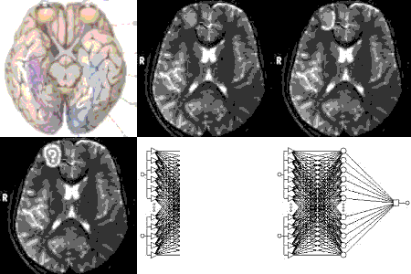
The purpose of that work is to automate the process of image analysis and conclusion about tumor by instrumental neural network implementation (figure 1.1).
Problems
Requirements to the program
Program have has the possibility to do the followings operations:
of image processing:
-to change contrast and brightness;
-to define the area of interest;
-to calculate parameters of area (size, density);
of conclusion results:
-to create database with necessary information of conducted inspection;
-to teach neural network for the purpose exposure of pathology;
-to conclusion of pathology type according to the tumor classification.
The format of programming processing is “*.jpg”. Microsoft Visual Basic 6.0 is chosen as the programm language.

Figure 1.1 - Animation (finding the tumor)
(11 shots, 7 cycles of repeating)
1.3 Scientific novelty and practical value
Use of neural networks and medical image processing
will allow to create effective specialized computer system. The program realized in master's work is unique in its way and
does not have analogues.
REVIEW ON THE TOPIC
2.1 Local and national review
Masters of DonNTU conducted developments for
such directions as the analysis of methods of contours selection, filtration, changes of image brightness,
teaching neural networks and etc.
Magnetic-resonance tomography is a method of reflection, used, mainly, in medical options,
for the receipt of high-quality images of human body's organs . MRT is based on principles
of nuclear-magnetic resonance, method of spectroscopy, used scientists for the receipt of
the molecules given about chemical and physical properties [4].
Developments of leading firms
Magnetic-resonance tomography «Gyroscan Intera»
firm «Philips» (Donetsk diagnostic center).
Information from magnetic-resonance tomography (images) can be saved in the format "*.DICOM",
and also in the format "*.JPG". Information from tomograph is passed to the doctor's computer and Easy
Vision station (Windows NT 4.0 operation system) [5].
Also there are such kind of systems:
- MRT System GE Signa Profile 0.2T
- MRT System Signa 1.5 Т MR/I [6].
The followings leading firms – producers of МRТ and CТ:
- MRT “ЭЛЕКТОМ-С5 (0.5 Tl)” - НИИЭФА the name of Efremov, Petersburg
- МRТ-0,02 (0.02 Тл) Kazan Physical-Technical Institute
- 2. МRТ “ИМТТОМ” (0.23 Tl) , firm "ИМТ-service", Moscow
DiViSy System
The system of DiViSy IP21 gives users the followings basic possibilities:
- Change of brightness
- Change of contrast
- Normalization of image in accordance with the histogram of distributing of brightness
- Inversion of brightness (a negative is a positive)
- Increase of sharpness of image (built-in filter of sharpen)
The followings
filters are realized in the current version of the system:
· Blur (smoothing)
· Prewitt 3 x 3 vertical (selection of vertical scopes)
· Prewitt 3 x 3 horizontal (selection of horizontal scopes)
· Sobel 3 x 3 vertical / horizontal
· Laplacian 3 x 3, 5 x 5
· Gaussian 3 x 3, 5 x 5 [7]
There is a system of medical image processing "Diamorf".
2.2 World review
Workstations “Easy Vision” and “Leonardo”
These workstations are widely used in tomography.
Functions:
- Computer angiography;
- high-surfaces reconstruction of three-dimensional pictures;
- three-dimensional reflection of volumes and surfaces;
- three-dimensional reflection of body underlying structures (Endo 3D) is in the interactive mode [4],[ 8], [9].
REVIEW OF METHODS
Because an initial image can contain different
noises, the program has possibility of image filtration (use of median filter). Median filtration is a method of nonlinear
signal processing. This method is useful for noise suppression of image. A median filter is a sliding window engulfing
the odd number of display elements. A central element is replaced by median of all display elements in a window. The
median of discrete sequence for odd N is such element, for which exists (N-1) /2 elements, less or equal to it on a size,
and (N-1) /2 elements, large or equal to it on a size. Median filtration represses separate impulsive hindrances effectively,
however can result of signal loosening [10, p. 342-346], [11, p. 123-128].
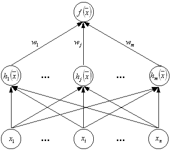
As an instrumental the neural network with the radial basic functions is chosen (figure 1.2).

Figure 1.2 – RBF network structure
The components of teaching set are:
- results of image processing;
- anamnesis information (complaints of patient);
- results of other inspections and analyses.
In RBFN as bases is chosen the functions of distance between vectors. The Euclid birth-certificate is used
as a measure of closeness. A neural network consists of three layers: input, hidden, output.
In other words, each one of n components of the input vector is passed to the inputs of m basis
functions and their outputs are linearly summed with the weights. The units of hidden layer obtains
the Gaussian function (formula 1.1).
 (1.1)
(1.1)
Thus, the RBF network output is linear combination of some set of basis functions (formula 1.2).
 (1.2)
(1.2)
The advantage of such kind neural networks is in their rapid teaching [12].
RESULTS
For realization of master's work the most optimum methods are chosen for medical image processing.
The area of interest is selected by doctor. This computer system is based on a neural networks design.
The neural network is creating and it will have the ability to react on input information and provide the diagnosis
of brain's tumor according to classification.
The program is developing. The full version will be in December, 2007.
CONCLUSION
The followings conclusions are made:
- There are many systems of image processing.
- Among the considered systems the most informing for tumor diagnostic is magnetic-resonance tomography.
- The image processing is made at the «Easy Vision» workstation of MRT.
- There is a possibility of automation of process of calculation all necessary parameters in area of interest.
- Operation of image processing and creation of the diagnosis system will be realize in master's work.
- There is a possibility to create the new improved diagnostic system which
will base on medical image processing and neural network teaching.
There is no similar system at the Donetsk Diagnostic center. Therefore I consider that this programm product will find the
application at the department of computer and magnetic-resonance tomography.
LITERATURE
- Журнал "Здоров'я України" http://www.health-ua.com/
- Электронная библиотека http://lib.chistopol.ru
- http://www.elinahealthandbeauty.com/
- Сайт о магнитно-резонансной томографии http://www.cis.rit.edu/htbooks/mri/contents-r.htm
- EasyVision, Philips Medical Systems, release 5.x, Nederland. – 2001
- Страница о разновидностях томографов http://www.stormoff.com/foreign/yamr.htm
- Группа компаний DiViSy является разработчиком и производителем профессиональных видеоконференций DiViSy для дистанционной работы в различных областях бизнеса, образования, медицины, экологии, производства, науки и т.д.
http://www.divisy.ru/
- Современное медицинское оборудование http://www.med-invest.ru/main/catalog/144/145/146/
- Сайт Донецкого диагностического центра (ДОКТМО) http://www.doktmo.donetsk.ua/ds/ds_01_kt.html
- William K. Pratt Digital image processing. – A Wiley-Interscience Publication, 1978
- Gonzalez, Rafael C. Digital image processing. – Prentice Hall, 2002
- Страница о нейронных сетях с радиальными базисными функциями http://www.basegroup.ru/neural/rbf.htm
To beginning
|


