
Abstract
Содержание
- Introduction
- 1. Theme urgency
- 2. Goal and tasks of the research
- 3. The results obtained to the time of writing the abstract
- References
Introduction
Computer processing of medical images involves the processing of digital images by computers or specialized devices based on digital signal processors.
All medical images, regardless of how they get to belong to one of two groups: analog and digital images. Analog images contain information of a continuing nature (eg, X-ray or ECG). Digital images are usually obtained by a computer and are based on a matrix that is contained in its memory. Thus, digital image, as opposed to analog, are discrete in nature.
When computer processing of medical images to solve a wide range of tasks such as image enhancement, the calculation of clinically important quantitative parameters of multidimensional spectral analysis of signals, recognition and image compression [1] .
Optical topographic survey methods spine are widely used in the 70s of last century, especially in the diagnosis of his distortions. All these methods are based on processing the patient's back pictures taken at different angles with a special light. In the future, for the processing of images were used special computer program, which was the basis of the method of computer-optical diagnostics (short - CODE). To process the photos back to the patient a few poses, illuminated by vertical stripes of light at a certain angle. Computer processing of these images to determine the amount and spinal muscle tension, to identify abnormalities in the arrangement of the bones, in particular, the magnitude of scoliotic arcs. All these data can later be used by specialists for an accurate diagnosis and appropriate treatment. The main advantages of this method are, first of all, his complete harmlessness (comparable to conventional photography), as well as high accuracy and meaningfulness of the results (comparable to the x-rays).
In a survey by the method of computer-optical diagnostics of the patient is placed on the mounting platform back to the camera, while the upper body should be exposed. Next, the number of pictures: in the natural position (with your back relaxed), the "active" position (with the "straight" effort back), if necessary, staff can take pictures in other positions. After processing the data in the computer results are displayed in a graphical representation of the surface spins in the corresponding planes [2].
1. Theme urgency
In establishing the diagnosis and treatment of conduct of doctors increasingly rely on medical images, which provide the bulk of information about the patient and his disease. However, their presence is not enough, because it requires analysis and interpretation, and the resulting information is used, then, for the diagnosis, treatment and during subsequent treatment planning.
Recognition of pathological processes is one of the most important tasks of medical imaging. However, the task of automated diagnosis of pathological processes in these medical images are still far from being solved.
2. Goal and tasks of the research
Goal of this work is to conduct a theoretical analysis of modern display techniques in the study of images and the results of their treatment, a comparison of the basic segmentation algorithms and the definition of perspective for their subsequent use in the analysis of medical images.
Main tasks of the research:
- Generalization literature and conducting a theoretical analysis of the results of imaging techniques in the study of images and the results of their treatment, as well as the known algorithms.
- Definition features of these algorithms and the selection of the best for use in the analysis of medical images.
- Development of software for the automated determination of pathology.
- Conduct numerical experiments, a comparative analysis of the results, formulating conclusions.
Research object: system of analysis and processing of medical images.
Research subject: informational, methodological and progarammno-algorithmic support of this system.
3. The results obtained to the time of writing the abstract
Processing and image analysis consists of the following steps.
- Pretreatment. Preprocessing phase removes abnormalities associated with the system generating the image and reduces noise.
- Change contrast. Calculation of the image histogram creates a representation of the number of pixels for each gray level in the image.
- Segmentation. This phase imaging isolates its individual elements.
- Calculation parameters. Calculation of linear and volumetric parameters of anatomical structures.
- Izobrazheniy.Poluchennye Interpretation of the structure and parameters should be compared with known structures and classified [10].
First stage of this work is to create a software product that allows to produce a visual and, as a consequence, an approximate estimate of existing abnormalities of posture. The program includes the following features: the ability to upload images of straight lines (vertical and horizontal), the construction of medium-sized vertical lines define the distance between the lines, changing the color or line thickness, calculate the angle between the lines, scaling the image, copy it and work with text.
Mathematical model
To implement the program functions relating to the calculation of distances and angles, the line is represented by two points: the beginning and end. Accordingly, each point has the coordinate axis X and Y. This allows the use of standard formulas and methods for working with coordinates on the plane.
Equal distance between two vertical parallel lines equal to the modulus of the difference of their coordinates on the X axis.
coordinates for the construction of the center line are computed from the middle segment.
Determination of the angle between the lines is carried out according to well-known formula for finding the angle between two vectors, each of which is given by its starting and ending point.
Software implementation
Executable program has been developed by means of Microsoft Visual Studio 2008, using application-level programming language C#. The program interface is, at present, is a composite and consists of a main window and some controls. It is made in the standard windows-style applications, which facilitates its perception.
Describe how to programmatically implemented the basic functions of this application.
Determining the distance between the lines is carried out in several stages.
first obtain the coordinates of the start and end points consistently distinguished lines.
Standard Definition workspace shape is 100 pixels per inch. Accordingly, 1 inch = 25.4 mm = 2.54 cm Therefore, a pixel has to 0.0254 cm, which corresponds to the real distance on the screen.
Since determine the distance between the parallel vertical lines, the calculation formula will be equal to:
distance = Math.Abs((p1 [0]. X + p1 [1]. X) / 2 - (p2 [0]. X + p2 [1]. X) / 2) * 0.0254, where
p1 – an array containing the coordinates of the first line,
p2 – an array containing the coordinates of the points in the second line,
Math.Abs – the function of taking the module.
By assumption, task grid spacing is 5 cm. Rounded the distance between grid lines is 25 pixels. That means 25 pixels = 5 cm, so 5 pixels = 1 cmdistance2 = Math.Abs((p1 [0]. X + p1 [1]. X) / 2 - (p2 [0]. X + p2 [1]. X) / 2) / 5 – the distance between lines, taking into account the mesh size.
Determination of the angle between the lines is as follows:
double abcd = (p1 [1]. X - p1 [0]. X) * (p2 [1]. X - p2 [0]. X) + (p1 [1]. Y - p1 [0] . Y) * (p2 [1]. Y - p2 [0]. Y) – the calculation of the scalar product.
double ab = Math.Sqrt ((p1 [1]. X - p1 [0]. X) * (p1 [1]. X - p1 [0]. X) + (p1 [1]. Y - p1 [0]. Y) * (p1 [1]. Y - p1 [0]. Y)) – find the length of the first line (vector).
double cd = Math.Sqrt ((p2 [1]. X - p2 [0]. X) * (p2 [1]. X - p2 [0]. X) + (p2 [1]. Y - p2 [0]. Y) * (p2 [1]. Y - p2 [0]. Y)) – find the length of the second line (vector).
double angle = Math.Round (rad * Math.Acos (abcd / (ab * cd)), 2) – calculation of the angle between the vectors and the transfer of the angle measures from radians to degrees.
if (angle> 90.0) {angle = 180 - angle;} – output only acute or right angle.
Building midline between the two marker lines is implemented as follows:
this.addLine (Math.Abs (p1 [0]. X + p2 [0]. X) / 2, p1 [0]. Y, Math.Abs ??(p1 [1]. X + p2 [ 1]. X) / 2, p2 [1]. Y, this.CreationPenColor, CreationPenWidth).
Stages of user interaction with the program
Steps and the order of user interaction with the program as follows:
1. Loading photos in JPEG (the patient against the calibration screen) (Figure 1).
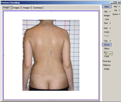
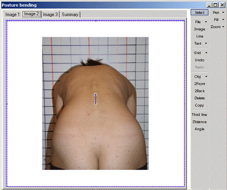
Figure 1 - Source images: photo 1, photo 2
2. In the images shown in photo 1 and 2, we construct three parallel lines. On the calibration screen to the right – it is 3 red lines – see Figure 2 and 3.
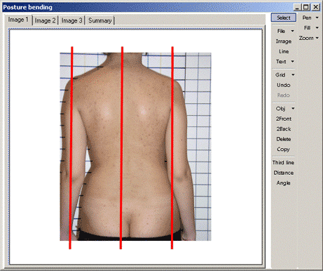
Figure 2 - Vertical marker lines on a photo

Figure 3 - Vertical marker lines on the photo 2
3. Held horizontal rib line (it passes through the highest point formed by the costal arch, parallel calibration lines) – Figure 5.
4. Calculate the deviation:
Photo 1:
- held the line on the gluteal fold;
- vertical lines are built along the contour of the neck;
- determined by the distance A, B, C (Figure 4).
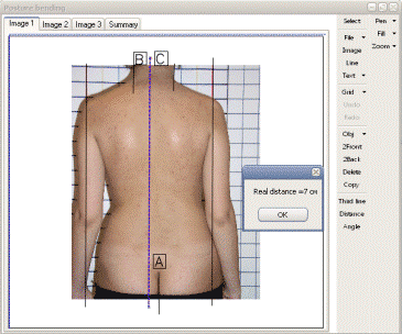
Figure 4 - Determination of the distance
Photo 2:
- held the line on the gluteal fold;
- construct a tangent line passing through the highest point formed by the costal arch;
- determined by the distance D and the angle E (see Figure 5).
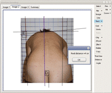
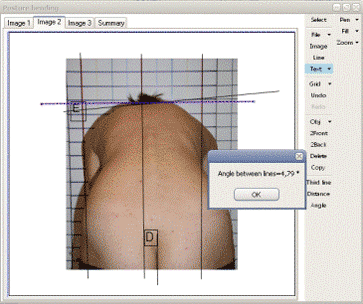
Figure 5 - Required construction
Thus, using the available tools in the program, you can make an automated assessment of the current patient's pathology.
Since the method is absolutely harmless, it can be used in surveys to assess the pathology and, consequently, to determine the suitability of the individual or the planning of training, depending on in what area and what age group will be applied.
This master's work is not completed yet. Final completion: December 2012. The full text of the work and materials on the topic can be obtained from the author or his head after this date.
References
- Панина А. И., Беловодский В. Н. Обработка медицинских изображений в среде разработки C#. NET.//Інформаційні управляючі системи та комп’ютерний моніторинг (КСМ, ІУС-2012) / Матерiали III мiжнародної науково-технiчної конференцiї студентiв, аспiрантiв та молодих вчених. — Донецьк, ДонНТУ — 2012.
- Панина А. И., Беловодский В. Н., Владзимирский А. В. Анализ медицинских изображений и диагностика патологии осанки в среде моделирования C#. NET.//Український журнал телемедицини та медичної телематики. — Донецьк, ДонНМУ ім. М. Горького, Науково-дослідний інститут травмотології та ортопедії — 2012, Том 10, № 1, с. 104-105.
- Sigurd Angenent, Eric Pichon, Allen Tannenbaum. Mathematical methods in medical image processing. – Bulletin (New Series) of the american mathematical society.
- Cufi X., Munoz X., Freixenet J., Marti J.A Review on Image Segmentation Techniques Integrating Region and Boundary Information [Электронный ресурс]. – Режим доступа: http://eia.udg.es/....
- Деткова Ю.Д.Обработка медицинских изображений с применением эволюционных алгоритмов в задачах биоинформатики. [Электронный ресурс]. – Режим доступа: http://img.avalon.ru....
- Виллевальде А. Ю. Система анализа и обработки медицинских изображений с малоконтрастными объектами : диссертация кандидата технических наук: 05.11.17 / Виллевальде Анна Юрьевна; [Место защиты: С.-Петерб. гос. электротехн. ун-т (ЛЭТИ)]. – Санкт-Петербург, 2008.– 143 с.: ил. РГБ ОД, 61 09-5/297.
- Близкая О. В. Разработка методов и алгоритмов обработки медицинских изображений с использованием методов искусственного интеллекта. [Электронный ресурс]. – Режим доступа: http://masters.donntu.ru ....
- Белявцев А. А. Разработка специализированной компьютерной системы диагностики клеток на основе анализа изображений. [Электронный ресурс]. – Режим доступа: http://www.uran.donetsk.ua ....
- Эль-Хатиб Самер Аднан. Компьютерная система сегментации медицинских изображений на основе алгоритма муравьиных колоний. [Электронный ресурс]. – Режим доступа: http://masters.donntu.ru ....
- Теоретический материал для подготовки к практическим занятиям и итоговому модульному контролю. Учебно-методическое пособие для студентов 2 курса медицинских вузов специальностей «Лечебное дело», «Педиатрия», «Стоматология». Под редакцией П.Е. Григорьева, Н.М. Овсянниковой. Авторы-составители: Овсянникова Н.М., Григорьев П.Е., Соколова Т.А., Ческая Т.Ю., Щеголева М.Г., Ислямов Р.И. [Электронный ресурс]. – Режим доступа: http://http://dmfi.info....