Dmitry Arkhypov
|
The abstract on a theme of masterful work
Abstract
Modern computer aids give wide functionality that allows to apply them in different areas. Introduction of mathematical methods, computer aids and new information technology, increase diagnostics possibilities in medicine where processing of the received data here and there and is conducted till now manually. An actual problem in the field of medicine is diagnostics and treatment of new growths of a liver. Presently the computer tomography is high on the list in the field of diagnostics of inwardness of the person. The most part of functional offices of hospitals use tomographs with which help it is possible to receive pictures of an internal. The subsequent processing of these pictures should be carried out to the highly skilled medical personnel. It attracts heavy expenses of time and means. Also not always processing and the inspection analysis is carried out in the necessary term for whatever reasons. Therefore automation of process of processing of such researches plays an important role in clinical practice. Necessity of creation of specialized computer system also consists in it.

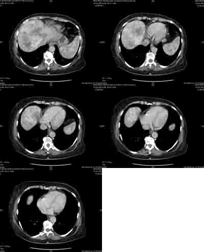
Fig1. Process of removal of tomograph of a liver.
(Animation, volume: 34kb, quantity of frames: 7, size: 200х164 рх)
For simplification of the task to doctors, and also the system of diagnostics is necessary for earlier diagnostics with the special software which will allow to define type of new growths. It will give possibility to the doctor more precisely to estimate the sizes of new growths, to accelerate treatment process. The system will improve quality of research, will give possibility quickly and conveniently to process the information, to keep account all patients, to store the diagnoses, the processed pictures, to supervise process to the even not not qualified expert, and besides doesn't demand the big economic expenses. Thus, working out SCS of the given type is an actual problem.
The purposes and work problems
1. A technique of carrying out of a computer tomography. Reception of the computer tomogram (cut) of a demanded site of a body at the chosen level is based on performance of following operations: - formation of the demanded width of a x-ray beam (collimation); - scanning of a demanded site of a body by a bunch of the x-ray radiation which are carried out by movement (rotary and forward) round a motionless body of the patient of the device "a radiator — detectors" - measurement of radiation and definition of its easing with the subsequent transformation of results to the digital form; - machine (computer) synthesis of the tomogram on a data set of the measurement, concerning the chosen layer; - construction of the image of an investigated layer on the monitor screen.
2. 2. Advantages of use of a computer tomography the Computer tomography possesses a number of advantages before usual radiological research: High sensitivity that allows separate bodies and fabrics from each other on density; the Computer tomography allows to receive the image of bodies and the pathological centers only in a plane of an investigated cut that gives the accurate image without stratification above and below lying formations; КТ gives the chance to receive the exact quantitative information on the sizes and density of separate bodies, fabrics and pathological formations that allows to do important conclusions concerning character of defeat; КТ allows to judge not only a condition of studied body, but also about mutual relation of pathological process with surrounding bodies and fabrics, for example tumor invasions in the next bodies, presence of other pathological changes; КТ allows to receive tomograms, i.e. The longitudinal image of investigated area like a x-ray picture by moving of the patient along a motionless tube. Tomograms are used for an establishment of extent of the pathological center and definition of quantity of cuts.
The review of existing methods of processing of tomograms Channelized researches is rather new and up to the end not developed. In the software products developed at present, processing of the image which is limited to a filtration, brightness and contrast regulation is carried out only. Thus the given software products don't carry and allocation of objects. All is the doctor is compelled to carry out manually that involves increase in time of processing of tomograms. I will try to solve this problem at the expense of creation of the software product capable by means of mathematical methods to allocate on the available tomogram objects, to define their parameters (the sizes, the area, number of objects, their brightness) and to assume, whether are these objects new growths and if yes that with what. One more important direction of researches in the field of processing of tomograms is creation of the automated systems of reception of the diagnosis on the basis of the given inspections and analyses. A problem here that algorithms of diagnosing of new growths aren't automated yet. Let's consider the basic methods of processing of the images, processings of tomograms used in existing systems. Filtration of the image on the basis of vejvlet-transformations
1. Selective veivelet
 Reconstruction of Vejvelet - the theory has been appreciably studied last years as the promising tool in compression of the image and noise reduction. In their works, Donoho and Johnstone developed theoretical structure discrete veivelet - transformations for an estimation of the signals deformed by additive white Gaussian noise. The principle, named selective veivelet - reconstruction is offered, and shown for reduction to optimum estimations for a wide variety of signals. We will assume that we have the deformed signal yi=xi+ni Gaussian noise N (0 σ2) Purpose consists in restoring the optimum appraiser xi for a desirable signal xi which results the minimum value of a square of an error. The scheme of selective vejvelet-reconstruction is illustrated on fig. 2. Drawing 2: the Block diagram for the scheme of suppression of the noise, based on discrete veivelet – transformation. The deformed image at first is transformed to a number veivelet - factors, that is, w=W (y) =? +z., where? And z - the factors corresponding to a desirable signal and noise. Process of threshold processing is applied further to veivelet - to factors, that is? =Тi (w), where t – threshold value. Sense of this approach - that the next pixels show high correlation which is translated only in slightly big veivelet – factors. On the other hand, noise is in regular intervals distributed among factors and in general the small. With correctly chosen value of a threshold noise can be effectively suppressed. Optimum value of a threshold t = σ 2log (N) (1) where N - the size of the block in veivelet - transformation.
2. Superfluous discrete veivelet – transformation As discrete veivelet – transformation not constant change, work on noise elimination could change considerably, changing initial moving, it also leads to some effects of blocks in the target image. The change constancy, however, can be reached calculation veivelet - transformations of all changes and performance of a threshold rule on each moved block. The received method name superfluous discrete veivelet – transformation. Algorithm following: 1. Execute noise elimination on the block of the size N on the basis of discrete veivelet – transformations. 2. Add the data without noise to corresponding position of the target image, and count number of the data for each sample. 3. Move a window horizontally and vertically, repeat a step 1 and 2 while all blocks in the image aren't settled. 4. Divide each input in the target image into number of repetitions. In our algorithm, we choose N=8, and we apply, or transformation of Haara (PH), or discrete transformation (DKP) as base of transformation. Strictly speaking, DKP not veivelet – transformation, it is chosen because of its good property of consolidation of energy so that the desirable signal is only in several positions in the transformed area, thus, leading to smaller artifacts of threshold processing. Experiments show that these two transformations spend comparable work. A firm and soft threshold rule both are applied. We will notice that the optimum size of a threshold for a soft threshold rule is received according to minimax criterion which means that possibility of improvement of work on noise elimination using various values of a threshold for specific images and transformations.
The mediannyj filter
The mediannyj filter unlike the smoothing filter realizes nonlinear procedure of suppression of noise. The mediannyj filter represents window W sliding across the field of the image covering odd number of readout. The central readout is replaced with a median of all elements of the image which have got to a window. A median of discrete sequence x1, x2..., xL for odd L name its such element for which exist (L - 1)/2 elements, smaller or equal to it on size, and (L - 1)/2 elements, big or equal to it on size. In other words, a median is average one after another a member of the number which is turning out at streamlining of initial sequence. Two-dimensional mediannyj the filter with window W we will define as follows:

As well as the smoothing filter, mediannyj the filter is used for suppression of additive and pulse noise on the image. Prominent feature mediannyj the filter, distinguishing it from smoothing, is preservation of differences of brightness (contours). Thus if differences of brightness bycicles in comparison with a dispersion of additive white noise mediannyj the filter gives smaller value СКО in comparison with an optimum linear filter. Especially effective mediannyj the filter is in case of pulse noise. As to pulse noise, that, for example, mediannyj the filter with a window 3х3 completely suppresses single emissions on a uniform background, and also groups of two, three and four pulse emissions. Generally for suppression of group of pulse hindrances the sizes of a window should be at least twice more sizes of group of hindrances. Among mediannyj filters with a window 3х3 the following is most extended:

Coordinates of the presented masks mean, how many time the corresponding pixel enters into the ordered sequence described above. A version mediannyj the filter is the method, overwhelming pulse noise and at the same time is minimum changing values of brightness on the initial image, consists in replacement of brightness of pixels of local maxima by local maximum value of brightness between borders and replacement of pixels of local minima by local minimum value between borders:

Here P (i) - initial intensity of a pixel i; P ' (i) - new value of intensity of a pixel i. The equation (1) represents a minimum from k pixels, the equation (2) - a maximum from k pixels. [7]
Detection of contours of object
For definition of characteristics of objects of the image it is preliminary necessary to separate them from a background, i.e. to find their borders. These borders represent curves on the image along which there is a sharp change of brightness or its derivatives on spatial variables. It is necessary to localize places of ruptures of brightness or its derivatives to learn something about the properties which have caused them of represented object. As edge is called the border between two areas, each of which has uniform brightness. The point is considered belonging to a contour if two conditions are simultaneously satisfied: 1. This point belongs to object; 2. This point has at least one next point which doesn't belong to object.
Conclusion
Generalizing works of the researchers who are engaged in processing of tomograms, it is possible to tell that for today the basic emphasis becomes on image improvement of quality, namely, filtrations. But thus the attention to search of objects on the image isn't paid. Process of manual processing of tomograms is now the big problem as leads to the big expenses of time and accordingly reduces throughput of offices of a computer tomography. In the work I have chosen the optimal methods filtrations from what are used in already existing systems of processing of tomograms. Using methods, I allocate objects on the tomogram, I define their sizes and the area. On the basis of the given approach, the computer system. The neural network capable adequately to react to entrance influences is created and to provide statement of the diagnosis of new growths of a liver that proves correctness of the chosen method. The realized software product will comprise experience and knowledge of leading experts in this area. Thus, use of neural networks at processing of computer tomograms of new growths of a stomach will allow to create effective SCS.
The list of references
1. Габуния Р.И. «Компьютерная томография в клинической диагностике»
2. Бобровнік Ю. «Сучасні програми постпроцесінгу та їх можливості»
3. Zeyun Yu, Chandrajit Bajaj «A fast and adaptive method for image contrast enhancement»
Исходный URL: http://ccvweb.csres.utexas.edu/cvc/papers/ICIP04.pdf
4. Зонневельд Ф.В. «Общая характеристика компьютерной томографии»
5. Лекции по обработке изображений http://graphics.cs.msu.ru/courses/cg02b/lectures/lection5/sld019.htm
6. Воскобойников Ю.Е. Касьянова С.Н., Кисленко Н.П.,Трофимов О.Е. «Использование алгоритмов нелинейной фильтрации для улучшения качества восстановленных томографических изображений»
7. Жирнов В.Т., Смирнов К.К., Трофимов О.Е. «О численных методах решения задач томографии»
8. Воскобойников Ю.Е. Колкер А.Б. «Адаптивный алгоритм фильтрации изображений и преобразования их в векторный формат»
9. Колкер А.Б. «Взвешенные и рекурсивные алгоритмы векторной медианной фильтрации»
