Zeyun Yu and Chandrajit Bajaj
Department of Computer Sciences, University of Texas at Austin, Austin, TX 78712-1188, USA
CONTENT
ABSTRACT
- INTRODUCTION
- ADAPTIVE CONTRAST ENHANCEMENT
- RESULTS
- CONCLUSION
ABSTRACT
In this paper we describe a fast approach for image contrast enhancement, based on localized contrast manipulation. Our approach is not only fast and easy to implement, but also has several other promising properties (adaptive, multiscale, weighted localization, etc.). We will also discuss in this paper an anisotropic version of our approach. Several examples of medical images, including brain MR images, chest CT images and mammography images, will be provided to demonstrate the performance of our approach.
1. INTRODUCTION
Many images, such as medical images, remote sensing images, electron microscopy images and even our real-life photographic pictures, suffer from poor contrast. Therefore, it is very necessary to enhance the contrast of such images before further processing or analysis can be conducted. There have already been many techniques for enhancing image contrast. The most widely used methods include various contrast manipulations and histogram equalization. Classic contrast manipulation is usually based on a globally defined stretching function (or called transfer function in the following). Histogram clipping might be needed before pixel-by-pixel stretching. Traditionally histogram equalization is also a global technique in the sense that the enhancement is based on the equalization of the histogram of the entire image. However, it is well recognized that using only global information is often not enough to achieve good contrast enhancement (for example, global approaches often cause an effect of intensity saturation).
To remedy this problem, some authors proposed localized (or adaptive) histogram equalization, which considers a local window for each individual pixel and computes the new intensity value based on the local histogram defined in the local window. The adaptivity can usually improves the results but it is computationally intensive even though there are some fast implementations for updating the local histograms. Furthermore, adaptive histogram equalization is a uniform local operator in the sense that all the pixels within the local window equally contribute to the determination of the new value of the center pixel being considered. Sometimes, like Gaussian filter, a weighted contribution of all the neighbors to the center pixel is more desired.
A more recently developed technique is called retinex model, in which the contribution of each pixel within the local window is weighted by computing the local average based on Gaussian function. A later version, called multiscale retinex model, gives better results but it is computationally more intensive. Another technique for contrast enhancement is based on wavelet decomposition and reconstruction and has been used for medical image enhancement, especially for mammography images.
In the present paper we propose a fast method for image contrast enhancement. The basic idea of our method is to design a transfer function for each pixel based on the local statistics. Our method follows the idea of the global contrast manipulation, but it also inherits the advantages of adaptive histogram equalization and retinex model. In addition, our method demonstrates a multiscale property, as shown later. First we describe the details of our approach. Then We present several examples of medical images and show the contrast-enhanced results by our approach. Finally we give the conclusion.
2. ADAPTIVE CONTRAST ENHANCEMENT
In this section we describe the details of our algorithm. In our method, a new intensity is assigned to each pixel according to an adaptive transfer function that is designed on the basis of the local statistics (local minimum/maximum as well as local average intensity).
2.1. Compute Local Min/Max/Avg
The local min/max/avg of a pixel can be simply defined as the minimal, maximal and averaging intensities within a local window of a fixed size. This is straightforward to implement but it has two problems. First, it takes a lot of time to search for the local min/max or compute the local average for each pixel. Secondly, the computed min/max maps always manifest some block-like artifacts. In the following we will compute the local min/max/avg maps using a propagation scheme.
One way to eliminate the block-like artifacts is to apply a Gaussian filter to the obtained min/max maps, yielding smoothed min/max maps. However, this requires additional time. The exponential and Gaussian filters can be implemented very fast by propagation scheme. This idea is directly applicable to the computation of the local average map. The propagation rule from a neighbor, say, (m - 1, n) to the pixel (m, n) is defined as follows:

where C is called conductivity factor, ranging from 0 to 1. The matrix lavg stands for the local average map, initialized with the image intensity values. The above propagation rule is sequentially applied in row & column directions. In order to compute the local min/max maps, we have to make some modifications on the above propagation scheme, we introduce in the following a conditional propagation scheme. Assume that lmin and lmax stand for the local min/max maps, respectively, and are initialized with the image intensity values. The conditional propagation scheme from (m - 1, n) to (m, n) is defined as follows:

2.2. Determine Transfer Function
Once we obtain the local statistics (local min/max/avg) for every pixel, we then need to design a transfer function from pixel to pixel. The essential idea for most contrast enhancement techniques is to take advantage of range stretching. In other words, the narrow intensity range seen in the original image is often expanded to a much broader range. In our method, the original range at a pixel is given by the absolute difference between the obtained local minimum and maximum intensities at that pixel, that is, |lmax - lmin|. This value is modified according to the curve illustrated in fig.1(a), where the x-coordinate represents the input range while the y-coordinate stands for the output range. This function curve is composed of two circular arcs:

where w0 is a fixed value. The threshold w0 is used such that, if |lmax - lmin|< w0, the contrast is thought of as noise and hence reduced. By this way we can suppress image noise while enhancing image features.

(a) Stretching window (b) Transfer function
Fig. 1. Determine the transfer functions
After the mapping from the original narrow range |lmax - lmin|, we obtain a broader range (denoted by w) such that lmin and lmax are mapped to 0 and w, respectively. In the meanwhile, the original image intensity Iold and average Aold, which satisfy lmin <= Iold, Aold <= lmax, must be linearly stretched to their new values Inew, Anew:

To achieve better contrast enhancement, we take account of the following observations. If the image intensity at a pixel is lower than the local intensity average, then we decrease the image intensity using a concave transfer function. On the other hand, if the image intensity at a pixel is higher than its local intensity average, we increase the image intensity using a convex transfer function. These transfer functions are adaptively defined from pixel to pixel, based on a parameter a, defined as: a = (Anew - Inew)/128. Specifically, we define the transfer function as a segment of parabolic curve, such that the transfer function is convex if a< 0 and concave if a > 0 (as seen in fig.1(b)). Note that those curves certainly could be in other forms but using parabolic curves makes it easier to derive a unified formula. To derive the explicit expression of those parabolic curves, we introduce a new coordinate system, namely, X'Y'-system (see fig.1(b)). In the new system, the parabolic curves can be represented as:

To derive the expression in XY-coordinate, we consider the following coordinate transformation:

Combining (4) and (5), we have an explicit expression:

where

After obtaining the transfer function for each pixel, we then compute the enhanced intensity as follows:

where f is the transfer function defined in (6).
2.3. Anisotropic Propagation
The conductivity factor C in (1) and (2) is a constant value. In term of filtering, however, this is isotropic, which may blur the features. A common technique to remedy this problem is known as anisotropic diffusion. With this idea in mind, we developed an anisotropic propagation for contrast enhancement. Eq.(1) and Eq.(2) become:

where R is called resistance factor and generally chosen from the interval [0.01,0.1] in our experiments.
3. RESULTS
We have tested our method on various types of images. In this section, however, we only show several examples of medical images and demonstrate the enhanced results. Fig.2(a) shows a brain MR image with low contrast. The enhanced image after applying histogram equalization and the result by our method (isotropic propagation with C = 0.95) are shown in Fig.2(b) and (c), respectively. Fig.3(a) shows an example of chest CT image. The enhanced image by histogram equalization and enhanced image by our method (anisotropic propagation with R = 0.1) are shown in Fig.3(b) and (c), respectively. Finally we demonstrate the multi-scale property of our method (by isotropic propagation) in Fig.4. The original image (Fig.4(a)) is a mammography image with very low contrast. Fig.4(b)(c)(d) illustrate results by our method using different conductivity factors, yielding different scales of image details that are enhanced.
4. CONCLUSION
In this paper we present a fast approach for image contrast enhancement. Our method is based on fast computation of local min/max/avg maps using a propagation scheme (either isotropic or anisotropic). We demonstrated the performance of our method and its multiscale property on three types of medical images. Our experiments on many other types of images also gave promising results both on computational speed and on enhancement quality.
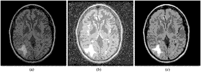
Fig. 2. Example 1: brain MR image—(a): original image with low contrast. (b): enhanced image by histogram equalization.
(c): enhanced image by our method (isotropic propagation with C = 0.95).

Fig. 3. Example 2: chest CT image—(a): original image with low contrast. (b): enhanced image by histogram equalization. (c): enhanced image by our method (anisotropic propagation with R = 0.1).
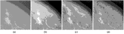
Fig. 4. Example 3: mammography image—(a): original image with very low contrast. (b): enhanced image by our method
with isotropic propagation (C = 0.95). (c): enhanced image by our method with isotropic propagation (C = 0.85). (d): enhanced image by our method with isotropic propagation (C = 0.75).
| 
 Мой e-mail: iluhin@sktel.com.ua