


Abstract of Master's thesis on the topic
1. Introduction
Currently, hospitals in vivo diagnostics of bone tissue in terms of mineral density by non-computer method - a standard linear rentgenodensitometry (rentgenofotometri). The optical density of images on rentenogramme was determined by densitometer or microphotometer, ie, devices intended for other purposes and adapted to the conditions of work. Such instruments and devices are based on the use of complex and expensive optical systems, which, moreover, are cumbersome. All this in the amount of obsolescence to some non-computer methods of diagnosis determines the urgency of this problem. A significant advantage of the projected SCS is no need to acquire a medical institution large number of complex and expensive equipment. Enough scanners to digitize x-rays.
2. Purpose and objectives
The purpose of master's work - designing specialized computer systems for the determination of bone mineral density by X-rays.
Projected SCS will undertake the following tasks:
- Initial processing of the digitized image in order to improve its image quality (removing artifacts, filtering, etc.).
- Recognition of the object image (the bones).
- Image recognition benchmark.
- Determination of the average brightness of the selected user image fragment of the object.
- Comparison of the average brightness of a fragment of the object image with a certain level of image brightness pattern.
3. Relevance and perceived scientific innovation
Preliminary assessment of the novelty of expected results shows the predominance of their practical value, so the spacecraft itself are processes, Methods and techniques realizatsiiopredeleniya bone mineral density is constant in the projected SCS with respect to existing non-computer systems, but the projected SCS has high practical value because of the widespread use of rengenoskopii in traumatology, orthopedics and other areas of medicine.
4. Review of existing developments
Currently, hospitals in vivo diagnostics of bone tissue in terms of mineral density by non-computer method - a standard linear rentgenodensitometriey (rentgenofotometrii). The optical density of images on rentenogramme was determined by densitometer or microphotometer, ie, devices intended for other purposes and adapted to the conditions of work. These devices contain complex and expensive optical systems, which, moreover, are cumbersome. All this in the amount of obsolescence non-computer methods of diagnosis determines the urgency of this problem.The system is the same sound in terms of medicine technique, which is the base for the developed my SKS. Unlike DEXA-scan system developed by SCS me is that the DEXA-scan does not use a traditional X-rays, more familiar to us will not print images on film. Output of X-ray machine at once arrive at the analog-digital converter, and digitally fed to a computer that is subject to further processing.
5. The list of outstanding tasks and deliverables
The study of mineral metabolism in the human body has attracted the attention of a wide range of experts in connection with mineral salts in the regulation of key functional processes. The most reliable method of determining the mineral saturation of bone ash content in the bone substance in vivo does not allow to establish the degree of mineral saturation of bone tissue. The methodology underlying the existing definition of bone mineral density by X-rays lies in the fact that X-rays, penetrating through the tissue, in this case bone, 95% delayed calcium phosphate and bone contains 98% of this chemical compound. Consequently, the degree of blackening of X-ray film is inversely proportional to the mineral saturation of bone tissue. The emergence of devices with an electrical recording of the optical density of blackening on the X-ray enabled with accurate register even small changes in the degree of blackening. In carrying out X-ray close to the study of the skeleton is placed on the cassette standard. As a result, the x-ray next to the bone image is obtained by the standard.
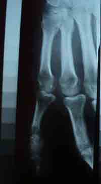
Figure 1. X-ray of the hand bones of man and an aluminum step-graded standard
After the photo processing of x-ray film using a photometer and densitometer the optical density image (mg/mm2) and investigated the site of bone and compared with the same optical density ipsAgen graded standard, which shows the density of the object in mg / mm ?. The ratio of the resulting density and the thickness of the bone gives an indication of bone density, that is, its mineral richness in mg/mm3. Using standard aluminum due to the fact that aluminum has a uniform structural composition (bone does not have a uniform crystalline structure), and empirically found that the graphic standards of aluminum provides more even and therefore more precise timetableThan the standards of other materials (the bone of cattle). The analyzed portion of the object image corresponds to the brightness with the brightness of the closest reference plots. Know in advance what portion of which corresponds to the standard mineral density. Simultaneous "photographing" the object and reference addresses a problem that the object and reference will be imprinted with different errors. The input data are the SCS projected digitized X-rays of any part of the musculoskeletal system of human. Imprint projected SCS - values of the average normalized optical density of the image of sampling sites of the bone.
Contrast:
Low contrast - the most common photographic, scanner, and television images, due to limited range of reproducible brightness. By contrast, is usually understood the difference between the maximum and minimum brightness values. By digital processing of the contrast can be improved by changing the brightness of each pixel and increasing the range of brightness. To this end, developed several methods. You can improve the contrast by using the normalization of the histogram. At the same time to fly a maximum range of brightness levels [0, 255] stretch is not the whole histogram, which lies in the range from fmin to fmax, and its most intense portion (fmin ', fmax'), are excluded from consideration uninformative "tails". The purpose of histogram equalization (this procedure is also called linearization and equalization equalization) is a transformation that, ideally, all levels of brightness would get the same frequency, and luminance histogram would correspond to a uniform distribution law.
Image filtering:
Real images, along with useful information include various obstacles. Interference sources are inherent noise of photodetectors, grainy photographs, the noise channels. Finally, the possible geometric distortions, the image may be out of focus. When processing of raster images, which consist of individual pixels, the integration is replaced by summation. The easiest way to implement the finite size of the PSF in the form of a rectangular matrix format of N N. N may be 3, 5, 7, etc.. To eliminate the effect of blurring the contours in the suppression of noise to make the transition to the nonlinear processing. An example of a nonlinear filter for noise suppression is the median filter. When median filtering (i, j)-th pixel is assigned the median value of brightness, ie, a value whose frequency is equal to 0,5.
6. Conclusion
Integration of modern computer technologies in medicine contributes to the fact that the computer diagnostic systems are widely used in medicine, and thus there is a wide range of options for automation research. Develop specialized computer system for determining bone mineral density does not create a revolution in Traumatology and Orthopedics. Doctors, as before, will perform radiographic examination of the patient, and the projected SCS will provide them an opportunity to accelerate and facilitate the process of examining X-rays. Scope projected SCS - any facility or department, specializing in trauma, orthopedics, as well as carrying out diagnosis of mineral metabolism (the level of osmotic pressure, the conduction of excitation in the nervous system, the automatism of the heart muscle, etc..) In the human body.
7. Literature
- Physics of imaging in medicine: In 2 vols. Volume 1: Trans. with English Ed. S. Webb. - M.: Mir, 1991. - 408.
- Physics of imaging in medicine: In 2 vols. Vol.2: Trans. with English Ed. S. Webb. - M.: Mir, 1991. - 423.
- Pratt W. Digital Image Processing: In 2 vols. Volume 1: Trans. from English. - Moscow: Mir, 1982. - 312s.
- Pratt Y. Digital Image Processing: In 2 vols. Vol.2: Trans. from English. - Moscow: Mir, 1982. - 325s.
- Ponomarenko, SI Pixel and vector. Principles of digital imaging. - St. Petersburg: BHV-Petersburg, 2002. - 496.
- Soros Educational Journal. 1996, № 2. Soifer VA Computer processing of images. Part 1.
- Soros Educational Journal. 1996, № 3. Soifer VA. Computer processing of images. Part 2.
- Newman, W., Newman, M. Mineral metabolism of bone. - M., 1961.
- Vaynshenker GA change in the degree of bone decalcification on radiographs. "Orthopedics and traumatology», № 2, 1967.