
Blood - that's life. Blood is the connecting link between all the organs and body systems. Blood parameters clearly reflect the functioning of the whole organism. That is why in medicine, a blood test is the primary research required to treat any disease. Circulating through the vessels in the body, blood supplies oxygen to tissue cells, which is necessary for the successful oxidation-reduction reactions and energy production. Dissolved nutrients, as well as protective factors are transported with blood. It maintains a constant temperature of our body. Regulation of all the systems functioning is carried out by blood. Besides analyzing the chemical and physical composition of blood by receptors, the human brain receives information about the state of the organism as a whole. Thus, blood is involved in maintaining the constancy of internal environment in the rigid framework. But what is the origin of blood? Despite the fact that it's still pretty vague question, at present it is believed that all blood cells come from the only one original cell - the parent polypotent cell that gives rise to various types of cells and can reproduce itself. Red blood cells make up the bulk of blood cells. They define the red color of blood. The number of red blood cells is normally 4 - 5 million in 1 mm 3 of blood for a healthy adult. Red blood cells - highly specialized cells, whose structure is subject to the fulfillment of their main function - to transport oxygen.
The role of cells in the body is diverse, primarily because of their specificity in different tissues and organs of the body. However, the general properties of the vast majority of cells are the vital processes such as attachment, phagocytosis, transformation, proliferation, necrosis, apoptosis. Observation of cells in the process of their life is the most important step to study their behavior. At the same time observation of the process of phagocytosis, attachment, transformation, proliferation, apoptosis and necrosis is difficult, since all cells are in constant motion, they are in huge number, and finding of the exact cell, which is currently in a particular state, is difficult. In real-time monitoring of a particular process of cell activity from its beginning to the end is difficult. For such reasons, modeling is a useful tool for studying cellular activity in real time, which requires development of a specialized computer system. Performance of this task requires 3D simulation environment, and consequently, a computer program product, which may make 3D modeling of objects. Many options of such software (Alias Maya 7.0, CyberMotion 3D Designer 10.0, Inivis AC3D 4.0.8, LightRay3D 1.3.4) are considered, but nevertheless, the most optimal and convenient environment modeling three-dimensional graphics program was chosen «3Ds Max», developed by the company «Autodesk». «3Ds Max» provides the ability to portray objects more accurately and perform various actions over these objects, which later can be saved as a video. This software was also selected because it includes a mechanism for calculating the physics and allows to simulate the behavior of hard and soft bodies, taking into account the gravity and other effects [3]. The program «3Ds Max» is easily accessible to any user of "the Internet", because it is distributed free of charge in the CIS.
The purpose of my research is to create a software product that under certain input parameters allows to model a definite (user selected) process of cell activity. The result of simulation is expected to see the video (animation) of the behavior of cells from the beginning the process to its completion. In order to simulate anything, one should have the information and data about the object model. Therefore, for this study, we need the geometrical parameters of blood cells (volume, area, radius of the section), and the parameters of the intercellular space (the pH level, temperature, ionic composition of the medium). These parameters are necessary for calculation of the cells vital processes rate, as well as for modeling more accurately of the behavior of realistic cell in the cellular environment. Analyzing the results of the program, it will be possible to make conclusions about the blood sector, or the whole human bloodstream. In this way, diagnosis and conclusions can be obtained much faster and more precisely by computer modeling. Quality program is useful for many medical institutions, will benefit them and save multiple physical resources and time.

Attachment – – the process by which the cell is attached to the surface and contacts with it closely and can lie prone. Schematically, the attachment is shown in Fig. 2. This process is the main and the first in the life of cells, because only after the attachment the cell begins to grow, eat, breed, etc. [4]. Initially, the cell is in suspension above the substrate (Fig. 2, a), then it slowly moves to the substrate and is attached to it (Fig. 2, b). The cells gradually spread on the substrate (Fig. 2, c).

Phagocytosis - - process in which a specially designed blood cells and tissues of the body (phagocytes) capture and digest infectious agents, and dead cells [4]. Phagocytosis is schematically depicted in Fig. 3. Initially, the cell is attached to the plane, for the implementation of any process of living cells are possible only after its attachment to the substrate (Fig. 3, a). Cell (phagocyte) absorbs bacteria: cell membrane envelops the bacterium and thus the bacterium is inside the cell (Fig. 3, b). Next is the splitting of the trapped object. (Fig. 3, c). Thus, the cell is fed.

Proliferation - cell proliferation (formation of new cells) [4]. Schematically, the proliferation is shown in Fig. 3. The cell nucleus is a doubling of DNA (Fig. 4, b). It produces trans ¬ river wall (Fig. 4, c). Then the daughter cells apart (Fig. 4, d). Thus, the cell multiplies. After finishing of proliferation daughter cells tend to move in one of two stationary states: either they re-enter the cell cycle (autosinteticheskoe state), or leave it (geterosinteticheskoe state). Cells in the resting state can exist in two states: G0 and G2. In the state of G0 DNA content in the cell is 2c, and able to G2 - 4c. Irreversible exit from the cell cycle is called the terminal differentiation.

Transformation - changes in the hereditary properties of the cell as a result of mutations [4]. Schematically, the transformation is shown in Fig. 5. Cell changes its unpredictable geometric parameters (Fig. 5, b, c).

Necrosis - pathological form of cell death, which results in cell swelling, increasing in size and collapsing [4]. Necrosis is schematically depicted in Fig. 6. Geometric parameters of the cell increase so that the cell membrane ruptures and the contents of the cell goes into the extracellular area (Fig. 6). Dead cells completely stop functioning. Cell death is accompanied by irreversible biochemical and structural changes.

The factors leading to necrosis: mechanical: effects on cells, tissues and organs of the mechanical force exceeding the resistance of shell, leading to their crush, tear, etc.; thermal: temperatures above +60 ° C or below - 15 ° C causes burns, frostbite ; electric: at entry and exit areas of the body of electric high tension develops very high temperatures, there are "signs of power" on the body - burned in those areas of tissue, chemical: strong acids, proteins coagulate cells, causing dry necrosis.
Apoptosis - form of cell death, manifested by a decrease in the size and decay into smaller parts that are absorbed by phagocytes [4]. Apoptosis is schematically shown in Fig. 7. The cell shrinks, and breaks up into fragments apoptotic cells (Figure 7, d). Morphologically, apoptosis is manifested loss of single, randomly placed cells, accompanied by the formation of rounded, surrounded by a membrane cells ("apoptotic bodies").

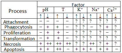
As it is known, the parameters of the intercellular spaces influence the behavior of cells. Effect of change in some environmental factors on the course of the cells vital processes are shown in Table. 1. The table is based on the article [2].
Recent research in this area is a scientific article by Gerasimov IG, Privalov, MV Grinchenko A. "Predicting the parameters of the process of attachment of blood cells in vitro» [1]. A specialized computer system is developed to predict the parameters of the process of blood cells attachment in the sample of neutrophils on the basis of kinetic model building process. Snapshot images of neutrophils obtained by using the ASUS Live Version 4.6 B2. With the help of a specially written program outlining and the calculation of the perimeter of neutrophils were performed. Parameters of the kinetic equation of the process of attachment, the calculation of average value and confidence interval were obtained using a statistical analysis package R for Windows FAQ Version for R? 2.4.1. Of the outstanding moments of this work it should be made that prediction is only for a single process - attachment and other processes are not considered. In addition, the software allows to work with ready-made images (pictures) and enables the simulation process.
The scheme of specialized computer system is developed to simulate the processes of attachment, phagocytosis, transformation, proliferation, apoptosis and necrosis of cells and to display the vital processes of cells gradually from beginning to end. Modeling allows to specify the original geometric parameters of the cell, as well as the parameters of the environment, which affects the speed of the process. With the real understanding of the process of blood cell activity it will be possible to draw conclusions about the blood general properties and circulatory system condition of the body.
This work is not completed yet. Final completion: Dec. 1, 2011. Full text of the work and materials on the subject can be obtained from the author after that date.