|
|
Faculty: Computer Information Technologies and Automatic Chair: Electronic Technique Specialty: Electronic Systems Subject of the master’s work: Research and substantiation of flow diagram of oxymeter by method of simulation modeling The supervisor of studies: с.t.s., doc. Hlamov |
|
|
~~~~~~~~~~~~~~~~~~~~~~~~~~~~~~~~~~~~~~~~~~~~~~~~~~~~~~~ Abstract The purpose and problems of work Prospective scientific novelty Existing methods and development Planned and received own result In medicine of critical statuses the important place occupies tracking indicators of function of external breath for the purpose of the control of process of gas exchange between an organism and environment. There is a necessity for creation of devices of diagnostics for an determination of authentic degree of the control o working capacity of lungs. To control concentration of oxygen in arterial blood is necessary of patients under a narcosis and in reanimation chambers. There are not enough devices for the operative control of the oxygen maintenance in blood for today. Pulsioxymetry is the most widespread, but this method gives the big error. There is a problem of more exact control of saturation of arterial blood oxygen. Controllable indicators of saturation are determinate saturation and partial pressure of oxygen. Normal indicators should be in limits of 95-98 % and 90–115мм hg. Therefore working out devices of the operative control of the maintenance of oxygen in blood is an actual prolem. The purpose and problems of work. The purpose of work: to make a model of the device of the operative control by concentration of oxygen in arterial blood. Measurement of percentage of oxygen and its control in blood hemoglobin are necessary at the control of viability of the patient during operation and in the course of restoration. Work problems is the substantiation of the photometric method based on fixation of the reflected stream, development and a substantiation of the structure, elements of the device, and also increase of accuracy of measurement and speed of the device. Prospective scientific novelty Scientific novelty consists in increase of accuracy by direct introduction of a photostream in a artery through special catheter, use of the reflected component of a stream, the account of such influencing factors, as pH of blood and a body temperature of the person. Existing methods and development There are three methods of definition of concentration of oxygen in blood: 1. Laboratory – demands invasive a capture of the sample of blood and does not give possibility to investigate concentration in a dynamic mode. For research needs very thin layer of blood, therefore in blood adds plasma that reduces accuracy of definition of oxygen. 2. Gasohromatographic – it is carried out by means of special chromatograph. The capture of test of blood, about 0,1-0,2 ml which is investigating within several minutes. Advantage of a method is high accuracy – ±3 %, however gas- chromatograph can be used only in laboratory, and also use of reactants represents inconveniences and brings an additional error in result of measurement. 3. Photometric – a method pulsioximetry. The technique of pulsioxymetry is based on used principles photopletizmography, allowing to allocate an arterial component of absorption of light for definition an oxygen in arterial blood. Measurement of this component gives the enable of using spectrophotometry for noninvasive monitoring oxygen saturation in arterial blood. (figure 1). In this case the signal from the data unit exit, proportional to absorption of light which is passing through fabrics, switches on two components: pulsing component, caused by change of arterial blood volume at each reduction, and the constant "base" component defined by optical properties of a skin, venous and capillary blood and other fabrics of an investigated section.
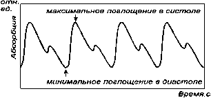
Figure 1 – The data unit of pulsioxymeter By the analysis of the form of a signal photopletizmogram (figure 2) it is possible to allocate its fragments corresponding to the moments of systolic emission. In these short time intervals at systole top it is possible to define most precisely saturation of oxygen in arterial blood.
Figure 2 – Photopletizmogram peripheral pulse For definition of saturation two-beam spectrophotometrical method is used. Measurement of absorption of light is made during the moments systolic emission, when signal has the maximum of amplitude. For this purpose in the data unit are used two sources of radiation with various spectral characteristics.
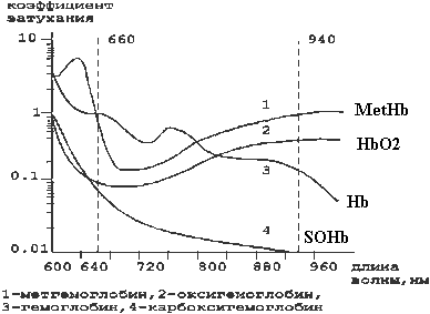
For definition of the greatest sensitivity of oxygen saturation is
necessary to choose lengths of waves of radiation of sources in
sectors of a spectrum with the greatest difference of light
absorption oxyhemoglobin and hemoglobin (figure 3). To this
condition satisfies red and infra-red areas of a spectrum of
radiation. At length of a wave of radiation of 660 nanometers (red
area) hemoglobin absorbs approximately in 10 times more light, than
oxyhemoglobin, and on a wave of 940 nanometers (infra-red area) -
absorption oxyhemoglobin more than hemoglobin. For measurement of
concentration of all four forms of hemoglobin is necessary to make
measurements of absorption of light, at least, on four lengths of
waves.
Figure 3 - Dependence of absorption of light on length of a wave of radiation for various forms of hemoglobin Pulsioxymeter is consists of the peripheral data unit, the microprocessor, the display showing a curve of pulse and value of saturation. The structure of pulsioximeter is shown in figure 4. The majority of devices have a sound signal of the certain tone which height is proportional of saturation that is very useful if the display pulsioximeter is not visible. The data unit is established in peripheral departments of an organism, for example, on fingers, an ear or a noses wing. In the data unit there are two light-emitting diodes, one of which radiates visible light of a red spectrum (660 nanometers), another – in an infra-red spectrum (940 nanometers). Light passes through fabrics to the photodetector, thus the radiation part is absorbed by blood and soft fabrics depending on concentration in them of hemoglobin. The quantity of absorbed light of each of lengths of waves depends on degree of oxygenation of hemoglobin in fabrics.
Figure 4 – The structure of pulsioxymeter Advantage of a method is instant reception of result, and deficiency is small accuracy and a considerable quantity of influential factors (temperature, bright light, vibration, etc.) elimination of which is connected with complication of the device or at all it is impossible. Hence, laboratory methods provide sufficient accuracy, but roads, big dimensional and cannot be used for the operative control over indicators. Photometric methods can be used for the operative control, but have low accuracy and depend on many influential factors. Planned and received own result As a result of work it is planned to develop the device for the operative control of blood’s saturation, providing accuracy in several percents. As blood dense enough substance at light passage through blood test the considerable part of a luminous stream is reflected. Therefore it is more expedient to define reflexion factor, instead of absorption factor. In case of the two-componental absorbing environment of measurement is necessary to make on two lengths of waves of optical radiation. As the basic length of a wave takes equal wave of 0,805 microns on which factors of absorption of hemoglobin and oxyhemoglobin are equal (see figure 3). For this case: c (l2) L = 4 p×g(l2) ×L/l2 = WL [k0 (l2) C0 + kg (l2) Cg] = W×L×k (l2) (1) Where c(l2) – absorption factor on the wave-length - l2; γ (l2) – factor of extinction on the wave-length - l2; L – length of an optical way of absorbing substance; k0 (l2) =kg (l2) =k (l2) – absorption factors of oxyhemoglobin and hemoglobin on the wave-length - l2; С0 and Сg – relative concentration oxyhemoglobin and hemoglobin; W –general weight of hemoglobin. From the last follows: W = 4 p×g(l2) / [k (l2)× l2] (2) As the basic measuring length of a wave takes the wave of radiation l1 = 0,66 microns on which factors of absorption of hemoglobin and oxyhemoglobin as much as possible differ. Taking into account previous, for a wave l1:
Taking into consideration, that С0 + Сg = 1, relative concentration oxyhemoglobin is defined as
Here values of factors of extinction on the wave-lengths l1 and l2 is defined by results of measurements of the reflected streams of radiation on these lengths of waves. Reflexion factors are calculated as the relation of the reflected stream to the falling stream on a surface of the absorbing environment. Calculation of values of factors extinction is carried out by the formula:
Where n – the factor of refraction of plasma of blood,
Structurally the device consists of optical and electronic unites. In the scheme of the optical unit is included special catheter, which entered directly into a pot and connected to sources and the receiver by means of an optical fiber (figure 5).
Figure 5 - Animation of the work of optical unit of the device
As a result of use of new circuit decisions and algorithms of
simulation it was possible to raise accuracy and efficiency of
control.
2. Булатов М.И., Калинкин И.П. . Практическое руководство по фотометрическим методам анализа -5-е изд., перераб.- Л.:Химия, 1986. - 432 с. Устранение влияния мешающих веществ специальными приемами фотометрических измерений 4. К.Ю. ЗюСоЧун Влияние различных факторов на кислородный баланс организма пациента во время операции. www.rusanesth.com/Genan/St_11_6.htm 6. Литвинова А.В., «Разработка и обоснование структуры прибора оперативного контроля содержания оксигемоглобина в крови человека»http:/masters.donntu.ua/2006/kita/litvinova/diss/index.htm7. Е.Хилл, М.Д. Стоунхэм, Оксфорд, Великобритания, Практическое применение пульсоксиметрии.8. Прикладная оптика: Учебник для оптических специальностей вузов. / М.И. Апенко, А.С. Дубовин, Г.В. Дурейко и др.; Под общ. ред. А.С. Дубовина, – 2-е изд., перераб. и доп. – М.: Машиностроение, 1992. – 480 с. 9. Скоков И.В. Расчет спектральных интерференционных приборов. – М.: Машиностроение, 1983. – 79 с., ил. – (Б-ка приборостроителя). 10 Имитационное моделирование в задачах оптического дистанционного зондирования / Креков Г.М., Орлов В.М., Белов В.В. и др. – Новосибирск : Наука, Сиб. отд-ние, 1988. – 165 с. 11. Джон Г.Вебстер «Медицинские приборы. Разработка и применение», К.:Медторг, 2004.-620с. 12. Климков Ю.М. Основы расчета опто-электронных приборов с лазерами. – М.: Сов. радио, 1978. – 264 с. 13. Методы цифрового моделирования и идентификации стационарных случайных процессов в информационно-измерительных системах / А.Н. Лебедев, Д.Д. Недосекин, Г.А. Стеклова, Е.А. Чернявский. - Л.: Энергоатомиздат.Ленинградское отделение, 1988.- 64 с. 14. Бендат Дж., Пирсол А. Измерение и анализ случайных процессов. – М.: Мир, 1974. – 464 с. 15. Stoneham MD,Saville GM,Wilson IH.Knowledge about pulse oximetry among medical and nursing staff.Lancet 1994:334:1339-1342 Ссылки на внешние источники действительны на 15.05.2009г. |


 (4)
(4)