Pic. 1. Sevet main types of microplasts.
Animation consist of 7 frames with 0.5 second delay, full delay is 3,5 seconds.

Introduction
Osteosintez - surgical replicon of bone splinetков through different fixative constructions, providing the protracted removal of their mobility. A purpose of osteosynthesis is providing of the stable fixing of splinet in correct anatomic position with the maintainance of functional axis of segment, stabilizing of area of break to the complete union. A method is to one of anchorwomen at treatment of breaks of jaws. As fixings plates, made from materials, possessing a biological, chemical and physical sluggishness, are usually used. All transferred properties are possessed by titan. The choice of titanic plates is made on the basis of layer pictures of spiral computer tomography [3].
The correct preliminary location of titanic plates will allow to minimize probability of complications and will result in reduction of time of operation. The correct choice of form and bend of mini plate will allow to save the regular anatomic shape of bone.
General raising of problem
Osteosintez is used in those cases, when unsurgical methods did not give the desired result or when it was exposed after the inspection of patient, that other methods will not provide an adequate replicon and effective fixing of splinetков. For the leadthrough of osteosynthesis it is necessary to conduct the analysis of data, got by additional researches: radiographs or pictures of spiral computer tomography (SCT).
Computer tomography it is a method of non-destructive layer research of underlying structure of object, was offered in 1972 by Godfrey Khaunsfildom and Allan Kormakom, awarded for this development of the Nobel bonus. A method is based on measuring and difficult computer treatment of difference of weakening of x-ray photography radiation different on a closeness fabrics.
Computer tomography (CT) — in wide sense, a synonym of term is a tomography (because all modern tomography methods will be realized by a computer technique); in narrow sense (in which used considerably more frequent), synonym of term x-ray photography computer tomography, because exactly this method put beginning of modern tomography [2].
The result of leadthrough of SCT is an array of layer pictures with the fixed distance between them. Every picture is a cut of maxillufacial area and kept in the file of the special medical format - .dcm (Digital Imaging and Communications in Medicine).
Presently basic information, necessary for the leadthrough of osteosynthesis based on the analysis of radiographs and clinical inspection (direct inspection of patient a doctor), that is insufficient in most cases and during an operation it is necessary to change a preliminary decision. It is necessary to develop SCS of determination of morphological changes at osteosynthesis of jaws, for the further use a doctor.
The possible methods of renewal of damage are determined in created SCS, analysing the layer cuts of computer tomography. In the case of single transversal break it is necessary to execute the graphic replicon of parts in a regular anatomic shape. In the case of comminuted breaks it is necessary to recover the form of bone and it is correct to locate fragments, if it is possible.
In each of the transferred cases it is necessary to pick up the most suitable titanic plate, bending it so that it in exactness repeated the anatomic structure of bone in this place and to a full degree saved the proportions of person.
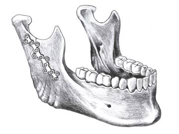
On a pic. 3 the example of single transversal break of lower jaw is resulted with displacement.
Comparison of wreckages and their fixing is needed in this place. For renewal of fragment it is necessary preliminary to project the supposed scopes of jaw in the site of damage, to confront parts in a regular anatomic shape. Then to define a type, form of titanic plate and place of its fastening. The result of SCS is passed to the doctor for consideration.
For the exposure of damages which need osteosynthesisе, it is necessary to process the cuts of SCT. By a file feature .dcm there is that an image and its size is kept in it, information about a patient and inspection, physical size of one pixel.
Mathematical methods
For determination of form of titanic plate it is necessary to take into account incurving of the damaged surface of jaw, the method of approximation splines is for what used. Whole part of jaw on either side appears a function which is determined in the site of fracture.
Under a spline usually understand the piece-set function, consilient with the functions of more simple nature on every element of breaking up of the range of definition.
The classic spline of one variable is built so: the range of definition is broken up on the eventual number of cuttings-off, on each of which a spline coincides with some algebraic polynomial. A maximal degree from the used polynomials is named the degree of spline. A difference between the degree of spline and turning out smoothness is named the bug of spline. For example, the continuous broken is spline of degree 1 and bug 1.
Splines have numerous applications both in a mathemati cal theory and in various calculable applications. In particular, splines two variables intensively used for the task of surfaces in the different systems of computer design.
Approximation splines is prognostication of passing of function in an interval wherever it is certain. For approximation use a cube spline [4].
Some function of f(x) is set on a segment [а,b], broken to pieces:
For the synonymous task of spline of the transferred terms not enough, for the construction of spline it is necessary to impose some additional requirements.
A natural cube spline is name a cube spline, satisfying also to the scope terms of kind:
Construction
We will designate: hi = xi − xi − 1
On every segment [xi − 1,xi] a function of S(x) is polynomial of the third degree of Si(x), the coefficients of which are necessary to be defined. We will write down for comfort of Si(x) in a kind:
then
The terms of continuity of all derivatives to the second order are inclusive written down in a kind:
and terms of interpolation are in a kind:
From here we get formulas for the calculation of coefficients of spline:

If to take into account that c0 = cn = 0, that calculation with it is possible to conduct by a method tuning-ups for a three-diagonal matrix [4].
A natural cube spline is name a cube spline, satisfying also to the scope terms of kind:
In developed SKS of determination of morphological changes for osteosynthesisе of jaw segmentation will be used.
Segmentation is a process of division of digital representation on a few segments (great number of pixels, also urgent superpixels). The purpose of segmentation consists in simplification è/èëè change of presentation of image, that it it was simpler and it is easy to analyse. Segmentation of images is usually used in an order to select objects and scopes (lines, curves, and ò. of ä.) on images. More exactly, segmentation of images is a process of appropriation of such marks to every pixel of image, that pixels with identical marks have general visual descriptions.
The result of image segmentation is a great number of segments, which cover all image, or great number of contours, abstracted from an image together. All pixels in a segment are alike by some recommendation or calculated property, for example in color, to the brightness or texture. Nearby segments considerably differ by this recommendation.
For the exposure of the damaged area it is necessary preliminary to execute image segmentation on results which it is possible to expose a break. The method of segmentation was chosen with the use of histogram, which is very effective, by comparison to other methods of segmentation of images, because it require an only one passage-way on pixels. In this method a histogram is calculated on all pixels of image and its minimums and maximums are used, to find objects dark-and-light.
Pictures of CT are images with prevailing black and former colors or close to them. We will build the histogram of distributing of brightness for a pic. 3.
Analysing a histogram it is possible easily to select objects dark-and-light, which are presented by single tops. For implementation of segmentation it is necessary to choose the threshold value of brightness, which. For every image this value is individual.
We will make segmentation of pic. 3 on the threshold of brightness 50.
This method by the best appearance allows to select objects dark-and-light and prepare it for a further analysis.
It is assumed in a prospect, that developed SKS of determination of morphological changes at osteosynthesisе of jaw, will visualize results which are passed for consideration of doctor for the further use.
Review of research-and-developments on the topic
Conducting the review of research-and-developments on the described topic, it was exposed following:
1. Global review.
It was not discovered analogues of the developed software which decides this or alike problem. The program which only exposes absent dark-and-light objects (bone, organ, soft fabric etc) and not engaged in their replicon of «Mimics» was found.
Mimics, there is processing of images software for the 3D planning and design, developed Materialise NV. Mimics creates and changes the surfaces of 3D-моделей drawing on the results of such обследовагний as a computer tomography (KT), to the confocal microscopy, микро-КТ and magnetic-resonance tomography (MRT) through the image of segmentation. 3D-файлы are presented in STL format. The most widespread format is an entrance of DICOM, but also other graphic formats, such as: TIFF, JPEG, BMP and raw material, supported also.
DICOM (Digital Imaging and Communications in Medicine) is standard for treatment, storage, print and transmission information in medical images. It plugs in itself the format of determination and network of protocol of connection. Protocol of connection of application uses TCP / IP for an exchange by information between the systems. DICOM files can be changed between two persons which are able to get images and information about patients in a format. DICOM allows to integrate scintiscanners, servers, work stations, printers and network equipment from different producers in archivings and transmissions of system images (PACS).
Formats of files of return to differ, depending on subsequent application: general 3D formats include STL, VRML, PLY and DXF. 3D-файлы can be optimized for FEA or CFD and can be exported in Abaqus in the format of INP.
2. National review.
It was now developed in Ukraine, to order of surgeons-stomatologies, software under the name «KORONIDA». This program is intended for a discovery and storage ground about a damage only of maxillufacial area, in future there is possibility to keep all stages of treatment.
The system of KORONIDA is a designer by which an user can expose and select the area of interest dark-and-light. Information in the system is presented as a chainlet of documents, reflecting all stages of treatment. The system is foreseen for work of both one user from a local computer and for simultaneous work of great number of users in a local network.
3. Local review.
It is not discovered on this query of results.
Conclusions
The mathematical methods described in the article will help to prepare an image for a further analysis, to expect the form of titanic plates.
Presently basic support at diagnostics of breaks of jaws is done on clinical examination. This SKS of determination of morphological changes at osteosynthesis of jaw will allow to automatize a process determination of factors, influencing on osteosynthesis.
Off-grade procedure of osteosynthesis can result in heavy complications. Qualification of doctor is very important therefore, observance by it all rules of leadthrough of procedure. Developed SKS provides exactness of positioning of titanic plate and decline of risk of postoperative complications. In spite of large experience of the use of mini plate in different countries, this method requires furher improvement.
Literature
1. Волков М. В., Гудушаури О. Н. и Ушакова О. А. Ошибки и осложнение при лечении переломов костей, М., 1979; — 216 p.
2. Соков Л. П. Курс травматологии и ортопедии, М., 1985; — 716 p.
3. Рычагов Г. П., Гарелик П. В., Кремень В. Е. Общая хирургия. — Мн.: Интерпрессервис; Книжный Дом, 2002. — 928 p.
4. Роджерс Д.,Адамс Дж. Математические основы машинной графики. — М.: Мир, 2001. — 340 p.
5. Костомаров Д.П., Фаворский А.П. Вводные лекции по численным методам. — 440 p.
6. Прасолов В. В. Задачи и теоремы линейной алгебры. — М.: Наука, 1996.— 342 p.
7. Linda G. Shapiro and George C. Stockman (2001): «Computer Vision», pp 279—325, New Jersey, Prentice-Hall. — 478 p.
8. Матрос-Таранец И.Н. Функционально-стабильный остеосинтез нижней челюсти. – Донецк, 1998. – 242 p.
9. Матрос-Таранец И.Н., Калиновский Д.К., Алексеев С.Б., Абу Халиль М.Н., Дадонкин Д.А., Челюстно-лицевой травматизм в промышленном мегаполисе: современный уровень, тенденции, инфраструктура - Донецк, 2001.- 193 p.
10. Vazirani V., Approximation Algorithms. — Berlin: Springer, 2003 — 284 p.
Important!
Master's work has not completed when this abstract was written. Completion of the master's work: December 2011. Full text of the work and materials on the topic can be obtained from the author or her supervisor after that date.